Short Communication
Intra Medullary Metastasis of Breast Cancer
1023
Views & Citations23
Likes & Shares
FINAL DIAGNOSIS
Intra Medullary Metastasis of breast cancer.
THREE DIFFERENTIAL DIAGNOSIS
Radiation myelitis -necrotizing myelitis - meningeal carcinomatosis.
IMAGE IN MEDICINE
INTRODUCTION
Intramedullary metastasis (MIM) is uncommon outcomes in breast cancer history [1,2]. They represent 1% of all metastases and 1 to 8% of the central nervous system (CNS) metastases [1-3]. Brain metastases and MIM are frequently correlated. The diagnosis is mainly based on medullary Magnetic Resonance Imaging (MRI) [4], showing the local tumor and distinguishing it from differential diagnosis, such as radiation myelitis, necrotizing myelitis or meningeal carcinomatosis.
CASE
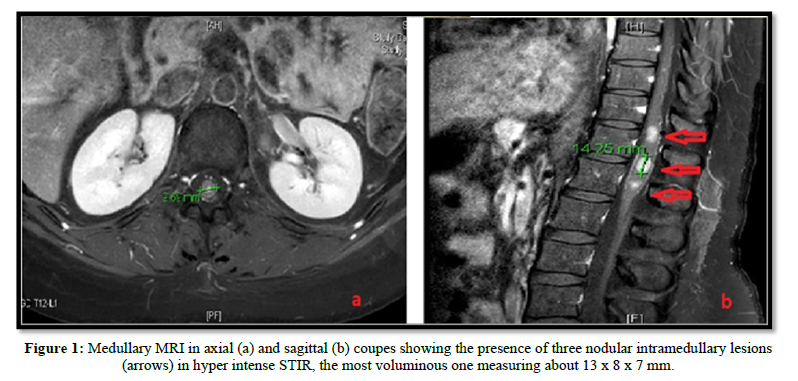
We report the case of a 35-year-old female patient, followed in Oncology-Radiotherapy department. Right breast biopsy revealed a grade 3 infiltrating ductal carcinoma. Hormonal receptors and Her-2 expression were found positive. Brain, liver and bones metastasis were detected at the time of diagnosis. She received brain radiotherapy (30 Gy) and then put under palliative oral chemotherapy associated with hormonotherapy with a good clinical outcome. Three months later, she developed neurological symptoms of medullary compression. The clinical examination found an incomplete left paraplegia with left leg anesthesia, right motor deficit (3/5). No sensory or sphincter disturbance were found and no pain symptoms. The MRI showed three nodular intra-medullary lesions localized at the terminal cone. The largest one was located on T12-L1, measuring 13 x 7 x 8 mm. The other two were located next to T12 and L1. The patient received high dose of corticosteroids and decompressive radiotherapy was performed with 30 Gy in 10 fractions of 3 Gy, improving initial symptoms. Another line of chemotherapy was recommended because metastatic disease progression (Figure 1).
DISCUSSION
Intramedullary metastases constitute a serious course of the cancerous disease [1-3].
It is rare and represents less than 1 to 2% of metastatic sites [4]. The localization at the cervical spinal cord is the most frequent [1-5]. Lung cancer is the primitive most often involved, followed by breast cancer which is frequent in our context, then comes melanoma, renal cancer and more rarely prostate carcinoma [1,3,5,6]. The hematogenous route is the most probable route of dissemination.
the diagnosis is essentially based on medullary MRI which most often shows nodular lesions in the medullary canal in T1 hypo signal - T2 hyper signal and after injection of Gadolinium [6].
the treatment is mainly palliative, requiring high dose corticosteroid therapy and radiotherapy on the affected marrow [7]. The evolution is often marked by a regression or a stability of the lesions but the prognosis remains poor because of the medullary localization and the speed of the evolution of the cancerous disease.
- Costigan DA, Winkelman MD (1985) Intramedullary spinal cord metastasis: A clinicopathological study of 13 cases. J Neurosurg 62(2): 227-233.
- Findlay JM, Bernstein M, Vanderlinden RG, Reach L (1987) Microsurgical resection of solitary intramedullary spinal cord metastases. Neurosurgery 21(6): 911-915.
- Bappon KD, Hirano A, Araki S (1959) Experiences with metastatic neoplasms involving the spinal cord. Neurology (Minneap) 9: 91.
- Schiff D, O’Neill BP (1996) Intramedullary spinal cord metastases: Clinical features and treatment outcome. Neurology 47: 906-912.
- Amin R (1999) Intramedullary spinal metastasis from carcinoma of the cervix. Br J Radiol 72: 89-91.
- Jardin F, Stamatoullas A, Fruchart C, D’ Anjou J, Glement JF, et al. (1999) Intramedullary spinal cord metastasis and leptomeningeal involvement in Hodgkin’s disease. Case report and review of the literature. Rev Med Intern 20: 267-271.
- Li Y, Takayasu M, Takagi T, Yooshimoto M, Mitsui Y, et al. (2000) Intramedullary spinal cord metastasis associated with hemorrhage: A case report. No ShinkeiGeka 28: 453-457.

QUICK LINKS
- SUBMIT MANUSCRIPT
- RECOMMEND THE JOURNAL
-
SUBSCRIBE FOR ALERTS
RELATED JOURNALS
- Ophthalmology Clinics and Research (ISSN:2638-115X)
- Journal of Immunology Research and Therapy (ISSN:2472-727X)
- Journal of Alcoholism Clinical Research
- Journal of Forensic Research and Criminal Investigation (ISSN: 2640-0846)
- Stem Cell Research and Therapeutics (ISSN:2474-4646)
- Dermatology Clinics and Research (ISSN:2380-5609)
- Journal of Clinical Trials and Research (ISSN:2637-7373)


