2587
Views & Citations1587
Likes & Shares
We describe 7 cases of retinal toxicity due to chloroquine in patients with systemic lupus erythematosus and drug use for 24 and 30 years, with doses of 250 mg daily that accumulate in total doses of 2190 g and 2737 g, respectively, for the first two cases and time of consumption between 2 and 6 years for the rest of the patients, with an average cumulative dose of 337.53 g. The patients underwent complete ocular examination, microperimetry, optical coherence tomography and multifocal electroretinography. The first case had a color vision dysfunction, without affecting visual acuity and contrast sensitivity, corneal deposits, loss of central retinal sensitivity of 14 dB in the microperimetry, evident fundus maculopathy, reduction of the retinal thickness in the macular area at 138 μm, with loss of the internal segment/junction of the external segment in the perifoveal region, loss of the foveal peak in the three-dimensional image with reduced P1 amplitude in the central rings in the multifocal electroretinogram. The second case showed a serious affectation of all the visual psychophysical parameters, diminished retinal sensitivity to 4 dB in the microperimetry, obvious maculopathy, macular thickness reduced to 108 μm, with loss of the internal/external segment in the macular, without foveal peak and P1 reduced Amplitude in all hexes in multifocal electroretinography. These cases illustrate different forms of retinal toxicity with chloroquine, apparently depending on the cumulative dose of the drug. The remaining 5 cases presented normal physical examination with slight loss of the internal/external segment of the photoreceptors, decreased retinal sensitivity in the microperimetry and decreased foveal peak in a smaller amount than the first two patients. Multifocal electroretinography, microperimetry and optical coherence tomography are useful to detect visual impairment at different stages of the disease.
INTRODUCTION
Chloroquine (CQ) is a 4 aminoquinolona used in the treatment of rheumatic diseases, particularly rheumatoid arthritis and lupus erythematosus since the early fifties. It can produce a dose-dependent iatrogenic affecting the retina, and was first described by Hobbs in 1959. Conducting research for the detection of retinopathy has been subject to controversy. Recent studies agree that it is unnecessary to conduct screening when the doses are below 3 mg/kg/day for a period of less than 5 years and there are no risk factors [1-4]. Currently the disease is still reported as a rare event, with a low incidence at least with current methods of screening.
In recent times perimetry fund or microperimetry has been used frequently for the functional assessment of patients with any type of maculopathy, using morphofunctional evaluation. Microperimetry accurately determines the location and fixation and stability of retinal threshold in the macular area, thus allows the projection medical diagnostic improve the various macular disorders.
We present two cases of chloroquine retinal toxicity after prolonged use, with different characteristics with regard to the pattern of damage and the characterization of the alterations by different diagnostic methods including microperimetry.
CASE REPORTS
2nd case
DISCUSSION
Retinopathy of chloroquine (CQ) is still reported as a rare event, especially after the increased use of hydroxychloroquine (HCQ) in the world, deriving the latter less toxic. In our environment by the limited availability of HCQ, CQ remains the choice in the treatment of rheumatic diseases with prolonged use due to the chronic course of the same. For these reasons it is more likely that patients taking this drug presented its toxic effects. The mechanisms by which toxic retinopathy occurs are not well known although it appears that oxidative stress caused anti-malarials in the retina play an important role. The CQ has acute effects on the metabolism of the cells of the retina, including photoreceptors. However, histopathologically, the earliest changes occur in the ganglion cell [5,6]. Clinically CQ retinopathy is characterized by bilateral and occasionally loss of visual field asymmetric shaped paracentral scotoma within 4 to 9 degrees of fixation and it precedes the development of clinical background, is often accompanied by defects in color perception. The first clinical data include a mild pigment mottling with loss of foveal reflex to evolve, in advanced stages, the typical target picture or bull's eye. The VA is compromised when there is macular involvement [5,7-9]. Alterations, if slight, can be reversed after discontinuation of medication. In advanced cases, the abnormalities may continue despite the suspension [2,5,10,11].
According to current trends despitaje studies for detection of retinal damage by consumption of CQ and its derivatives, there is a risk of toxicity with daily doses above 3 mg/kg of weight for more than 5 years of use and cumulative dose greater than 460 g. The elderly over 60 years and kidney disease, liver and retinal are also basic retinotoxicity potential risk [1,12,13]. Recently, the American Academy of Ophthalmology has updated its recommendations for the retinotoxicity pesquizaje by CQ and its derivatives, suggesting a series of diagnostic tools including multifocal electroretinogram (mfERG), optical coherence tomography in the frequency domain (SD-OCT), retinal autofluorescence and automated perimetry 10-2 program, all useful for their sensitivity in detecting early-stage retinal damage, which is really the goal of despitaje studies [1] As it is known that in advanced stages when maculopathy appears, it is highly unlikely the recovery of visual acuity.
The mfERG according to different studies seems to have become the gold standard test to detect CQ damage in early stages and to follow after discontinuing the drug [11,16-19]. In our patients despite being in different stages of toxicity with and without maculopathy, the mfERG demonstrated retinal dysfunction, affecting P1 amplitudes in all cases and changes in implicit time only in cases with bull eyes maculopathy. The rings 1 to 4 were the most affected, with major changes in the patient with a higher cumulative dose of the drug. Has been shown by other authors that the time of prolonged medication use and therefore higher cumulative doses predisposes to retinal toxicity and therefore changes occur gradually over the mfERG [16].
Microperimetry test is useful for the study of macular diseases and available in our environment in all ophthalmology centers. Allows a detailed study of central retinal sensitivity with accurate topographic correlation be-tween the fundus details. In our patients select the program Macula 20 degrees, 4-2 threshold strategy, Goldmann III. In the first case without maculopathy, the fixation was stable with a decrease in mean retinal sensitivity 6 dB with respect to normal values between 18 and 20 dB, despite not being impaired visual acuity and contrast sensitivity this study to quantify the functional deficit. The second case presented with an unstable fixation reduced the sensitivity to 12 dB compared to normal, functional impairment was higher, corresponding to structural changes. In the third and fourth cases, the fixation was stable with a decrease in the mean retinal sensitivity by 5 dB. The fifth case presented a decrease of 6 dB and the remaining two cases of 4 dB.
We are the criteria that microperimetry, allows an analysis of the visual quality reliable and reproducible, when examining the same retinal points in each study, which joined the mfERG that determines the focal retinal sensitivity and the changes that occur even before clinically evidencing the damage, are useful tools to allow monitoring with the same parameters evaluated in the initial examination. In the second case by the severe impairment of visual acuity, visual rehabilitation was started, for which the microperimeter MP1 was useful in the evaluation of fixation.
CONCLUSION
1. Gilhotra JS, Mitchell P, Healey PR, Cumming RG, Marmor MF, Kellner U, Lai T, Lyons J, Mieler W (2011) Revised recommendations on screening for chloroquine and hydroxychloroquine retinopathy. Ophthalmology 118: 415-422.
2. Browning DJ (2002) Hydroxychloroquine and chloroquine retinopathy: screening for drug toxicity. Am J Ophthalmol 133: 649-656.
3. Quijada E, Pareja A, Mantolán C, Cordoves LM, Losada MJ, et al. (2007) Screening protocol side effects of anti-malarials. Arch Soc Canar Oftal.
4. Marmor MF (2004) Current recommendations on screening for hydroxychloroquine or chloroquine retinopathy. Sociedad Iberoamericana de Información Científica (SIIC).
5. Rodríguez FJ (2000) Retinal toxicity and retinal pigment epithelium by chloroquine and hydroxychloroquine. J Rheumatol 7: 37-41.
6. Shroyer NF, Lewis RA, Lupski JR (2001) Analysis of the ABCR (ABCA4) gene in 4-aminoquinoline retinopathy: Is retinal toxicity by chloroquine and hydroxychloroquine related to Stargardt disease? Am J Ophthalmol 131: 761-766.
7. Jimenez-Palop M (2006) Malaria: an update of its use in rheumatic diseases. Rev Spanish Soc Rheumatol 2.
8. Orozco-Gomez LP, Ruiz-Morfin I (2005) Bull's-eye maculopathy. Rev Mex Oftalmol 79: 51-53.
9. Mavrikakis I, Sfikakis P, Mavrikakis E, Rougas K, Nikolaou A, et al. (2003) The incidence of irreversible retinal toxicity in patients treated with hydroxychloroquine. Ophthalmology 110: 1321-1326.
10. Lyons JS, Severns ML (2007) Detection of early hydroxichloroquine retinal toxicity enhanced by ring ratio analysis of multifocal electroretinography. Am J Ophthalmol 143: 801-809.
11. Gaynes BI, Torcznski, Varro Z, Grostern R, Perlman J (2008) Retinal toxicity of chloroquine hydrochloride administered by intraperitoneal injection. J Appl Toxicol 28: 895-900.
12. Restrepo JF, Guzmán R, Iglesias A (2002) As anti-malarial drugs induce remission in rheumatoid arthritis. J Rheumatol 9: 62-68.
13. Ferreras A, Pinilla I, Abecia E, Pajarin AB, Honrubia FM (2007) Retinal toxicity secondary to treatment with chloroquine. Arch Soc Esp Oftalmol 82: 103-108.
14. Swanson WH, Cohen JM (2003) Color vision. Ophthalmol Clin North Am 16: 179-203.
15. Rodriguez-Padilla JA, Hedges III TR, Monson B, Srinivasan V, Wojtkowski M, et al. (2007) High-speed ultra-high-resolution optical coherence tomography findings in hydroxychloroquine retinopathy. Arch Ophthalmol 125: 775-780.
16. Lai TY, Chan WM, Li H, Lai RY, Lam DS (2005) Multifocal electroretinographic changes in patients receiving hydroxychloroquine therapy. Am J Ophthalmol 140: 794-807.
17. Marmor MF (2005) The dilemma of hydroxychloroquine screening: new information from the multifocal ERG. Am J Ophthalmol 140: 894-895.
18. Elder M, Rahman AM, Mclay J (2006) Early paracentral visual field loss in patients taking hydroxychloroquine. Archiv Ophthalmol 124: 1729-1733.
19. Maturi RK, Yu M, Weleber RG (2004) Multifocal electroretinographic evaluation of long-term hydroxychloroquine users. Arch Ophthalmol 122: 973-981.
-
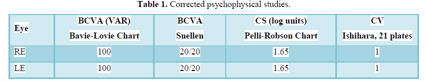
 Table 1
 -
Table 2
-
Table 3


QUICK LINKS
- SUBMIT MANUSCRIPT
- RECOMMEND THE JOURNAL
-
SUBSCRIBE FOR ALERTS
RELATED JOURNALS
- Stem Cell Research and Therapeutics (ISSN:2474-4646)
- Journal of Alcoholism Clinical Research
- Journal of Cell Signaling & Damage-Associated Molecular Patterns
- Journal of Clinical Trials and Research (ISSN:2637-7373)
- Journal of Spine Diseases
- International Journal of Surgery and Invasive Procedures (ISSN:2640-0820)
- Journal of Cardiology and Diagnostics Research (ISSN:2639-4634)


