Research Article
Study of Energy Biomarkers during Unpredictable Chronic Stress (UCS)-Induced Sprague Dawley Rats after Treatment with the Biofield Energy Treated/Blessed Proprietary Test Formulation
423
Views & Citations10
Likes & Shares
Unpredictable chronic stress (UCS) model was used to evaluate the effect of Consciousness Energy Healing/Blessing Treatment (the Trivedi Effect®) on a novel proprietary test formulation in male Sprague Dawley (SD) rats for the estimation of energy biomarkers by estimation of adenosine triphosphate (ATP) in brain, muscle, and liver along with stomach serotonin and citrate synthase activity using ELISA assay. A test formulation was formulated including minerals (magnesium, zinc, copper, calcium, selenium, and iron), vitamins (ascorbic acid, pyridoxine HCl, alpha tocopherol, cyanocobalamin, and cholecalciferol), Panax ginseng extract, β-carotene, and cannabidiol isolate. The constituents of the test formulation were divided into two parts; one section was defined as the untreated test formulation did not receive any treatment, while the other portion of the test formulation and three group of animals were received Biofield Energy Healing Treatment by a renowned Biofield Energy Healer, Mr. Mahendra Kumar Trivedi. The results showed that the level of ATP in brain tissue was altered as compared with the both disease control (G2) and untreated test formulation (G4) groups. Moreover, the level of ATP in liver was increased minimally in the G7 (15-days pre-treatment of Biofield Energy Treated Test formulation) and G9 (untreated test formulation to the Biofield Energy Treatment per se to the rats) groups, respectively as compared with the G4 group. Additionally, the ATP levels in the muscles was altered in all the treatment groups as compared to the G2 group. The level of serotonin in stomach was significantly increased by 30.8%, 44.8%, 91.2% (p≤0.05), and 36.3% in the G5, G6, G7, and G9 groups, respectively as compared with the G4. Besides, the expression of mitochondrial enzyme - citrate synthase in all the treatment groups was altered as compared to the both G2 and G4 groups. Overall, the energy biomarkers were significantly improved after Biofield Energy Treatment and could be beneficial for low metabolic energy disorders like Niemann-Pick disease, Tay-Sachs disease, Gaucher disease, Galactosemia, etc. The results also showed the significant slowdown the stress-related disease progression and its complications/symptoms in the preventive Biofield Energy Treatment group per se and/or Biofield Energy Treated Test formulation groups (viz. G6, G7, G8, and G9) as compared to the disease control group.
Keywords: Biofield Treatment, Energy Panel, ATP, Mitochondrial assay, The Trivedi Effect®, Unpredictable Chronic Stress, ELISA
INTRODUCTION
Depression is considered as a common neuropsychiatric disorder, which is reported to affect up to 20% of the world population [1]. Although, the symptoms may vary in terms of its presence and severity among individuals, however, in broad way it may include decreased energy, low mood and anhedonia, altered weight and appetite, sleep disturbances, irritability, and cognitive deficits, etc. [2]. In recent days, the researchers reported the emergence of a key component of the stress response i.e., mitochondria. These are subcellular organelles that help in sustaining life by taking part in the energy transformation and intracellular signaling [3,4]. The recent discoveries indicated the role of mitochondria in stress response at two major levels, i.e., as a target of stress; and as mediator of stress pathophysiology [5]. Besides, it was reported that the exposure to unpredictable chronic stress may induces an increase energy generation that could be further correlated with the increase in the production of reactive oxygen species (ROS). It is well known that the low levels of ROS production help in maintaining the physiological functions, such as, host defense, proliferation, gene expression, and signal transduction [6]. Mitochondria act as the main site for the generation of cellular ATP by the process of oxidative phosphorylation, in which there might be some leakage of the high-energy electrons in the respiratory chain during the process that may form the ROS. Under normal physiological conditions, the body maintains a cellular balance between the generation of ROS and its clearance, with the help of several antioxidative defense mechanisms, such as, enzymes and antioxidants [7]. For example, in the process of cold stress adaptation, the organisms increase the energy metabolism, which ultimately resulted in an overall increase in the capacity for ATP production as well as the increase of ROS generation. However, if there is the overproduction of cellular ROS that overwhelms the intrinsic antioxidant capacity, then it causes the oxidative stress, which may damage the biomolecules of normal cells as well as tissues. Moreover, the excess of ROS may also cause oxidative damage to the proteins, deoxyribonucleic acid (DNA), and lipids [8]. Besides, the exact mechanism behind the ROS increase in the body remains unclear, however, it is considered to be mediated in part by stress-related hormone such as cortisol [9].
It is known that the parts of the human body that are major oxygen consumers are muscle and liver, which might take up nearly 40% of cardiac oxygen delivery. Similarly, the brain is known for representing only 2% of body weight, but it takes nearly 20% of the total body oxygen. Therefore, the cellular homeostasis can be disturbed in the presence of oxidation-modified compounds and molecules, which may cause apoptosis or necrosis [10,11]. Also, such events are related to various neurodegenerative diseases as the neuronal cells present in the brain are highly sensitive to oxidative stress due to their greater dependence on oxidative phosphorylation for energy in comparison to other cell types [12-14]. ATP as a biomarker of energy panel, mitochondria assay, and serotonin were estimated as the standard biomarkers of energy level after inducing stress. In order to access the level of energy biomarkers, or change in level in different groups in presence of unpredictable chronic stress, a novel test formulation was designed. The test formulation was the combination of vital minerals (selenium, zinc, iron, calcium, copper, and magnesium), essential vitamins (cyanocobalamin, ascorbic acid, pyridoxine HCl, alpha tocopherol, and cholecalciferol), and nutraceuticals (β-carotene, Ginseng, cannabidiol isolate (CBD)). All the minerals and vitamins used in the test formulation have significant functional role to provide vital physiological role [15-17]. Besides, cannabidiol itself has wide range of pharmacological profile and was reported to role in different disorders [18,19], while ginseng extract is regarded as the one of the best immune booster and overall relaxing herbal medicine for overall managing immunity [20].
Biofield Energy Healing Treatment was reported to be beneficial against various pathological cases of immune disorders, stress-related diseases, aging, cervical cancer, and many more have found to be significantly beneficial. Biofield Therapy was accepted and defined as one of the best Complementary and Alternative Medicine (CAM) treatment [24-26]. National Center for Complementary/Alternative Medicine (NCCAM) recommended CAM with several clinical benefits as compared with the conventional treatment approach [27]. National Centre of Complementary and Integrative Health (NCCIH) accepted Biofield Energy Healing as a CAM health care approach in addition to other therapies such as deep breathing, natural products, Tai Chi, yoga, therapeutic touch, Johrei, Reiki, pranic healing, chiropractic/osteopathic manipulation, guided imagery, meditation, massage, homeopathy, hypnotherapy, special diets, relaxation techniques, movement therapy, mindfulness, Ayurvedic medicine, traditional Chinese herbs and medicines in biological systems [28,29]. The Trivedi Effect®-Consciousness Energy Healing Treatment was scientifically reported on various disciplines such as in the materials science [30,31], agriculture science [32], antiaging [33], gut health [34], nutraceuticals [35], pharmaceuticals [36], overall human health and wellness. The present study was aimed to evaluate the effect of unpredictable chronic stress (UCS) on the level of ATP in brain, liver, and muscles of male Sprague Dawley rats along with mitochondrial assay and estimation of stomach serotonin in the presence of the novel test formulation using standard ELISA assay, which was treated with Biofield Energy Treatment (the Trivedi Effect®) by a renowned Biofield Energy Healer.
MATERIAL AND METHODS
Chemicals and Reagents
Pyridoxine hydrochloride (vitamin B6), calcitriol, zinc chloride, magnesium (II) gluconate, and β-carotene (retinol, Provit A) were purchased from TCI, Japan. Copper chloride, cyanocobalamin (vitamin B12), calcium chloride, vitamin E (Alpha-Tocopherol), cholecalciferol (vitamin D3), iron (II) sulphate, and sodium carboxymethyl cellulose (Na-CMC) were procured from Sigma-Aldrich, USA. Ascorbic acid (vitamin C) and sodium selenate were obtained from Alfa Aesar, India. Cannabidiol isolate and Panax ginseng extract were obtained from Panacea Phytoextracts, India and Standard Hemp Company, USA, respectively. Imipramine hydrochloride was purchased from Sigma, USA. For the estimation of energy panel, mitochondria and serotonin estimation, specific ELISA kits were used such as for detection of ATP level and serotonin were procured from Bio vision and CUSABIO, USA, respectively.
Study Design
The current experiment was designed to fulfil the study protocol, animals were assigned into nine (9) groups. G1: Normal control; G2: Disease control (UCS: Unpredictable chronic stress + 0.5% CMC); G3: Reference item (UCS + Imipramine hydrochloride 30 mg/kg); G4: (UCS + Untreated test formulation); G5: (UCS + Biofield Energy Treated test formulation); G6: (UCS + Biofield Energy Treatment per se to animals from day -15; G7: (UCS + Biofield Energy Treated test formulation from day -15); G8: (UCS + Biofield Energy Treatment per se plus Biofield Energy Treated test formulation from day -15), and G9: (UCS + Biofield Energy Treatment per se animals plus untreated test formulation).
Maintenance of Animal
Randomly breed male Sprague Dawley (SD) rats with body weight ranges from 200 to 300 gm were used in this study. The animals were purchased from M/s. Vivo Bio Tech, Hyderabad, India. Animals were randomly divided into nine groups based on their body weights consist of 6 animals of each group. They were kept individually in sterilized polypropylene cages with stainless steel top grill having provision for holding pellet feed and drinking water bottle fitted with stainless steel sipper tube. The animals were maintained as per standard protocol of the Committee for the Purpose of Control and Supervision of Experiments on Animals (CPCSEA), Ministry of Environment and Forest, Govt. of India. The test facility is registered (registration no. 64/PO/br/s/99/CPCSEA) for animal experiments with the CPCSEA. The animals were procured using protocol approved by the Animal Ethics Committee (IAEC/41/505) and the husbandry conditions were maintained as per the recommendations of the CPCSEA.
Consciousness Energy Healing Strategies
Each ingredient of the proprietary test formulation was divided into two parts, one part of each was did not receive any sort of treatment and were defined as the untreated or control sample. The second part of the test formulation and three group of animals were treated with the Trivedi Effect® - Energy of Consciousness Healing/Blessing Treatment (Biofield Energy Treatment) by a renowned Biofield Energy Healer, Mr. Mahendra Kumar Trivedi under laboratory conditions for ~3 min. The Biofield Energy Healer was located in the USA; however, the test formulation was located in the research laboratory of Dabur Research Foundation, New Delhi, India. The energy transmission was done remotely to the test samples and animals. After that, the Biofield Energy Treated sample was kept in the similar sealed condition and used as per the study plan. In the same manner, the control test formulation group was subjected to “sham” healer for ~3 min under the same laboratory conditions. The “sham” healer did not have any knowledge about the Biofield Energy Treatment. The Biofield Energy Treated animals were also taken back to experimental room for further proceedings.
Experimental Procedure
Seven days after acclimatization, the animals were randomized and grouped based on the body weight. Dosing for groups G7 and G8 were initiated on day -15 and continued till end of the experiment. However, G1 to G5 and G9 groups were dosed from day 1 till the end of experiment. G6 group was not to be dosed with the test formulation. Body weight and clinical signs were taken daily throughout the experimental period. All the animals except G1 group received stress induced procedures such as sound stress, tilted cages and crowd stress, cold and warm water swim stress, food and water deprivation, stress due to change in the light and dark cycle were undergo seven different types of unpredictable stress procedures after scheduled dosing daily at specified interval to the end of the experiment for 8 weeks after the initiation of stress, which vary every week interval i.e., shuffling of stress type. During 8th week of the experimental period, all the animals were individually subjected for blood collection for the experimental purpose.
Preparation of Sample for ELISA Assay
With the continued stress treatment of 4th week of the experimental period, all the animals were individually subjected for blood collection using retro-orbital plexus and the blood was collected in the plain vial, which was used for the separation of serum in all the animals of different experimental groups. The serum from all the groups was stored at -20°C for further estimation. Alternatively, an aliquot of all the samples and store samples at -20°C or -80°C. Avoid repeated freeze-thaw cycles, which may alter the level of ATP in brain, muscle, and liver along with serotonin and mitochondrial assay during final calculations.
Estimation of Energy Panel (ATP), Serotonin, and Mitochondrial Assay
The serum from all the groups was subjected for the estimation of level of ATP in brain, muscle, and liver along with estimation of serotonin in stomach, and mitochondrial assay. The entire assay and the energy panel was estimation using ELISA method as per manufacturer’s recommended standard procedure. This was a quantitative method and the principle was based on the binding of antigen and antibody in sandwich manner assay.
Statistical Analysis
The data were represented as mean ± standard error of mean (SEM) and subjected to statistical analysis using Sigma-Plot statistical software (Version 11.0). For multiple groups comparison One-way analysis of variance (ANOVA) followed by post-hoc analysis by Dunnett’s test and for between two groups comparison Student’s t-test was performed. The p≤0.05 was considered as statistically significant.
RESULTS AND DISCUSSION
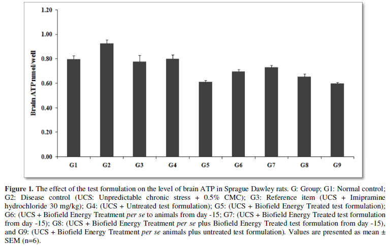
Estimation of ATP in Brain
ATP and the function of brain had a well coordination with respect to initiation of stress disorders [37]. Study suggested that the ATP level in brain was normally lower in case of the stressed or depressed patients as compared to the control subjects [38]. The stressed group animals were well correlated with the untreated and the test formulation treated groups. The level of ATP in all the experimental groups and is presented in Figure 1. The data suggested that unpredictable chronic stress group (G2) showed value as 0.93 ± 0.03 nmol/well, which was increased by 16.5% as compared with the normal control (G1, 0.79 ± 0.03 nmol/well). Positive control group, imipramine treatment (G3) showed decreased brain ATP (0.78 ± 0.05 nmol/well) by 16.1% as compared to the G2. Untreated test formulation to the untreated rats (G4) showed a decreased level of brain ATP (0.80 ± 0.03 nmol/well) by 13.7% as compared to the G2. Biofield Energy Treated test formulation to the untreated rats (G5) decreased brain ATP level by 34.1% and 23.6% as compared to the G2 and G4 groups, respectively. Biofield Energy Treatment per se to the rats (G6) showed significant decreased brain ATP level (0.69 ± 0.02 nmol/well) by 25.1% and 13.2% as compared to the G2 and G4 groups, respectively. However, 15-days pre-treatment of the Biofield Energy Treated test formulation (G7) significantly decreased brain ATP level (0.73 ± 0.01 nmol/well) by 20.8% and 8.2% as compared to the G2 and G4 groups, respectively. 15-days pre-treatment of the Biofield Energy Treated test formulation to the Biofield Energy Treatment per se to rats (G8) significantly decreased the brain ATP level (0.65 ± 0.02 nmol/well) by 29.6% and 18.4% as compared to the G2 and G4 groups, respectively. Similarly, the untreated test formulation to the Biofield Energy Treated rats (G9) showed significant decreased brain ATP level (0.60 ± 0.01 nmol/well) by 35.3% and 25% as compared to the G2 and G4 groups, respectively.
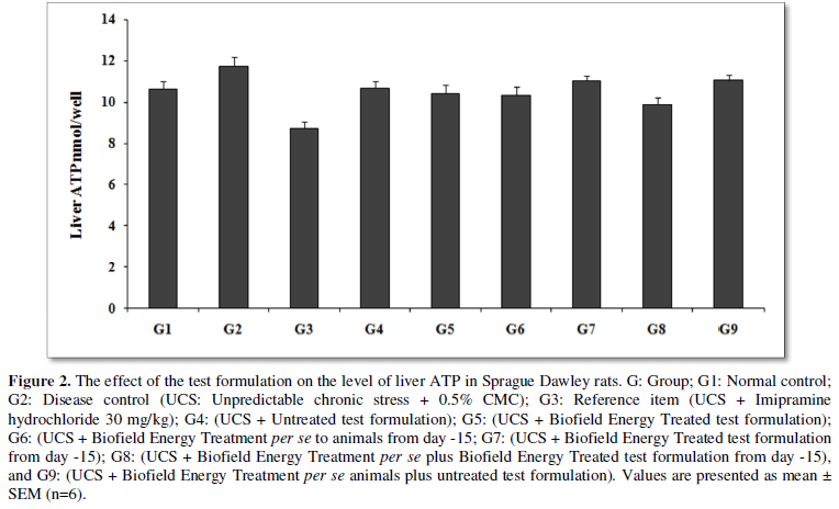
Estimation of ATP in Liver
Stress plays a vital role in liver disorders and main cellular energy source in liver is ATP. Liver disorders results in an altered ATP synthesis and liver steatosis [39, 40]. The effect of the test formulation was compared with respect to the untreated test formulation and Biofield Energy Treatment per se for energy panel in liver. ATP in muscle was estimated in presence of the effect of the test formulation, which was measured in all the experimental groups and are graphically presented in the Figure 2. The data suggested that unpredictable chronic stress group (G2) showed liver ATP level was 11.74 ± 0.4 nmol/well, i.e., increased by 10.3% as compared with the control (G1, 10.65 ± 0.4 nmol/well). Imipramine treatment (G3) showed decreased liver ATP level (8.74 ± 0.3 nmol/well) by 25.6% as compared to the G2. The untreated test formulation to the untreated rats (G4) showed decreased liver ATP (10.68 ± 0.3 nmol/well) by 9.1% as compared to G2. G5 group showed decreased liver ATP level (10.41 ± 0.4 nmol/well) by 11.4% as compared to G2. G6 group showed decreased liver ATP level (10.34 ± 0.4 nmol/well) by 12% and 3.2% as compared to the G2 and G4 groups, respectively. G7 group showed decreased ATP level (11.03 ± 0.2 nmol/well) by 6.1% as compared to the G2; while increased by 3.3% as compared to the G4 group. G8 group animals showed significantly decreased ATP level (9.89 ± 0.3 nmol/well) by 15.8% and 7.4% as compared to the G2 and G4 groups, respectively. G9 group showed decreased ATP level (11.06 ± 0.3 nmol/well) by 5.8% as compared to the G2; while increased by 3.5% as compared to the G4 group.
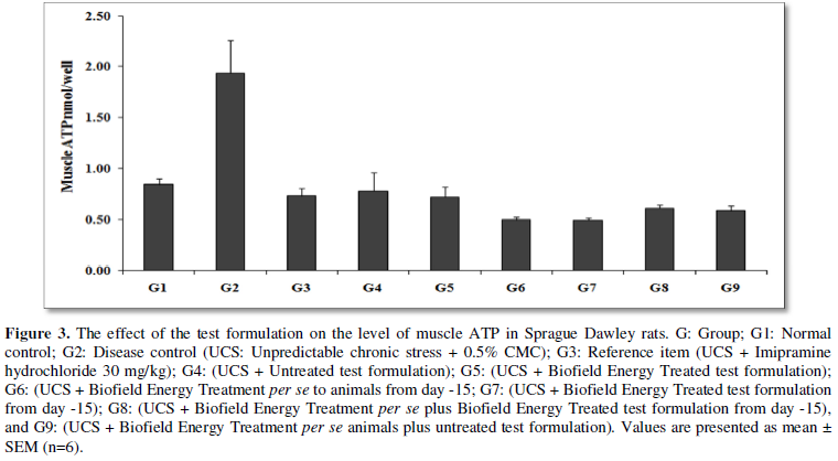
Estimation of ATP in Muscle
Muscle fatigue is one the common non-specific symptom-related with muscle ATP. ATPase has reported to be useful in management of tropomyosin for relaxation. Fatigue can be managed with the help of intracellular ATP. Muscular performance would be optimum in presence of muscle ATP and its energy [41,42]. The level of ATP in muscle was measured in all the experimental groups are graphically presented in the Figure 3. The data suggested that unpredictable chronic stress group (G2) showed muscle ATP level was 1.93 ± 0.3 nmol/well, i.e., increased by 128.9% as compared with the control (G1, 0.84 ± 0.1 nmol/well). Imipramine treatment (G3) showed decreased muscle ATP level (0.74 ± 0.1 nmol/well) by 61.9% as compared to the G2. The untreated test formulation to the untreated rats (G4) showed decreased muscle ATP (0.78 ± 0.2 nmol/well) by 59.4% as compared to the G2. G5 group showed decreased muscle ATP level (0.72 ± 0.1 nmol/well) by 62.5% and 7.6% as compared to the G2 and G4 groups, respectively. G6 group showed decreased muscle ATP level (0.50 ± 0.0 nmol/well) by 74.2% and 36.4% as compared to the G2 and G4 groups, respectively. G7 group showed decreased ATP level (0.50 ± 0.0 nmol/well) by 74.3% and 36.8% as compared to the G2 and G4 groups, respectively. G8 group animals showed decreased ATP level (0.62 ± 0.0 nmol/well) by 68% and 21.1% as compared to the G2 and G4 groups, respectively. G9 group showed decreased ATP level (0.59 ± 0.0 nmol/well) by 69.4% and 24.6% as compared to the G2 and G4 groups, respectively.
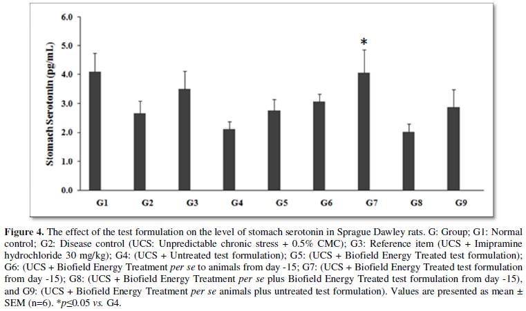
Estimation of Stomach Serotonin
Stomach and intestine are the primary reservoir of serotonin; it helps in controlling the bowel movements, function, regulate anxiety, happiness, and mood. Stress or stressful life events had a significant effect on gastrointestinal disease such as chronic disorders of the digestive system like inflammatory bowel disease (IBD), gastro-oesophageal reflux disease (GERD), gastrointestinal disorders (FGD), and peptic ulcer disease (PUD) [43]. Serotonin, biogenic amine [5-hydroxytryptamine (5-HT)] functions as a neurotransmitter play a vital role in stress conditions [44]. Stomach serotonin level in the unpredictable chronic stress (G2) was 2.67 ± 0.4 pg/mL, which was decreased by 34.9% as compared with the normal control (G1, 4.10 ± 0.6 pg/mL). Imipramine treatment (G3) showed an increased stomach serotonin level (3.49 ± 0.6 pg/mL) by 31% as compared to the G2. The untreated test formulation to the untreated rats (G4) decreased stomach serotonin level (2.12 ± 0.3 pg/mL) by 20.5% as compared with the G2. G5 group animals showed an increased stomach serotonin level (2.77 ± 0.4 pg/mL) by 4% and 30.8% as compared to the G2 and G4 groups, respectively. G6 group showed an increased stomach serotonin level (3.07 ± 0.3 pg/mL) by 15.1% and 44.8% as compared to the G2 and G4 groups, respectively. G7 group animals showed a significant increased stomach serotonin level (4.05 ± 0.8 pg/mL) by 51.9% and 91.2% (p≤0.05) as compared to the G2 and G4 groups, respectively. G8 group animals showed decreased stomach serotonin level (2.03 ± 0.3 pg/mL) as compared to G2 and G4. G9 group animals showed an increased stomach serotonin level (2.89 ± 0.6 pg/mL) by 8.3% and 36.3% as compared to the G2 and G4 groups, respectively (Figure 4).
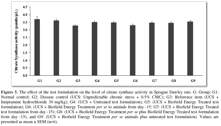
Estimation of Muscle Mitochondrial Assay - Citrate Synthase Activity
Mitochondria, play a major function in production of energy in cellular survival and death. Damage-associated molecular patterns (DAMPs) have been reported as a vital source in mitochondria. However, stress and its related factors causes major oxidative stress and inflammation that have been associated with cellular damage and citrate synthase activity, which is a main source of mitochondrial dysfunction [45]. The effect of the test formulation and Biofield Energy Treatment per se was estimated using the level of citrate synthase activity; the results are graphically presented in the Figure 5. Citrate Synthase activity in the unpredictable chronic stress (G2) was 3.63 ± 0.08 µunits/µL, which was decreased by 2.1% as compared to the normal control (G1, 3.71 ± 0.19 µunits/µL) group (G1). Imipramine treatment (G3) showed 3.27 ± 0.09 µunits/µL reduced level by 9.9% as compared to the G2. G4 group showed reduced value by 2.9% (3.52 ± 0.08 µunits/µL) as compared with the G2. However, group G5, G6, G7, G8, and G9 showed slight reduced value of Citrate Synthase level by 3.6%, 8.8%, 4%, 4.4%, and 2%, respectively as compared with the G2. Similarly, G5, G6, G7, G8, and G9 showed slight reduced value of Citrate Synthase level by 0.7%, 6.1%, 1.2%, 1.6%, and 0.9% respectively, as compared with the G4.

In this research plan, four groups were considered as preventive maintenance groups. These groups were G6 (Biofield Energy Treatment per se to animals at -15 days), G7 (Biofield Energy Treated test formulation from day -15), G8 (Biofield Energy Treatment per se to animals along with Biofield Treated test formulation from day -15), and G9 (Biofield treatment per se at -15 days to animals with untreated test formulation). The results showed the significant slowdown of the disease progression, stress-related all other symptoms/complications and also reduced the chances of disease susceptibility in these groups. Based on the overall data, it suggests that the Biofield Energy Healing/Blessing Therapy was found to be most effective and benefited in order to prevent and protect from the occurrence of any type of diseases in rat model. It indicated that this therapy can act as a preventive maintenance therapy to prevent the occurrence of the disease, slowdown the disease progression and disease-related complications of the existing aliments that will ultimately improve the overall health and quality of life in human.
CONCLUSION
The unpredictable chronic stress (UCS) animal model was tested experimental for estimation of energy biomarkers with special reference to ATP level in brain, muscle, and liver along with level of stomach serotonin and mitochondrial citrate synthase activity. The level of ATP in brain was estimated and compared with respect to untreated test formulation and other preventive measure groups and was altered. The ATP level in liver was increased in the G7 and G9 groups, respectively as compared with the G4. Additionally, the ATP levels in the muscles was altered in all the treatment groups as compared to the G2 group. In addition to, stomach serotonin level was significantly increased by 30.8%, 44.8%, 91.2%, and 36.3% in the G5, G6, G7, and G9 groups, respectively as compared with the G4. Further, the expression of mitochondrial enzyme - citrate synthase in all the treatment groups was altered as compared to the both G2 and G4 groups. Thus, Biofield Energy Healing Treatment (the Trivedi Effect®) per se and other preventive maintenance groups (G7, G8, and G9) showed outstanding results in rat model study. The energy biomarkers were significantly improved after Biofield Energy Treatment and could be beneficial for low metabolic energy disorders like Niemann-Pick disease, Tay-Sachs disease, Gaucher disease, galactosemia, etc. It also helped to slowdown the disease progression and disease-related complications of the overall animal’s health. These data suggested that Biofield Energy Treatment per se and/or Biofield Energy Treated Test formulation in combination would be the best treatment strategies in order to prevent and protect from the occurrence of any type of diseases. Therefore, the Biofield Energy Treatment/Blessing might act as a preventive maintenance therapy in order to maintain good health, or full restoration of health or improve the overall health and quality of life in human. This therapy might also reduce the severity of any type of acute/chronic disease (auto-immune related and inflammatory disorders) progression rate and can be used in both before and after the manifestation of any disease symptoms in healthy, unhealthy, and ill peoples such as many thyroid disorders such hyperthyroidism, goiter, thyroid cancer, Hashimoto's thyroiditis, etc. This test formulation also can be used against fibromyalgia, Addison disease, multiple sclerosis, myasthenia gravis, aplastic anemia, psoriasis, rheumatoid arthritis, Crohn’s disease, vitiligo, chronic fatigue syndrome and alopecia areata, as well as various inflammatory disorders such as ulcerative colitis, dermatitis, hepatitis, diverticulitis, mental disorders, Parkinson’s and other movement disorders, stroke and transient ischemic attack (TIA), and in the improvement of overall health and quality of life.
- Kessler RC, McGonagle KA, Zhao S, Nelson CB, Hughes M, et al. (1994) Lifetime and 12-month prevalence of DSM-III-R psychiatric disorders in the US. Results from the National Comorbidity Survey. Arch Gen Psychiatry 51: 8-19.
- Nemeroff CB (1998) The neurobiology of depression. Sci Am 278: 42-49.
- Morava E, Kozicz T (2013) Mitochondria and the economy of stress (mal) adaptation. Neurosci Biobehav Rev 37: 668-680.
- Picard M, Juster R-P, McEwen BS (2014) Mitochondrial allostatic load puts the ‘gluc’ back in glucocorticoids. Nat Rev Endocrinol 10: 303-310.
- Picard M, McEwen BS (2018) Psychological stress and mitochondria: A systematic review. Psychosom Med 80(2): 141-153.
- Dröge W (2002) Free radicals in the physiological control of cell function. Physiol Rev 82(1): 47-95.
- Cui H, Kong Y, Zhang H (2012) Oxidative stress, mitochondrial dysfunction, and aging. J Signal Transduct 2012: 646354.
- Nita M, Grzybowski A (2016) The role of the reactive oxygen species and oxidative stress in the path mechanism of the age-related ocular diseases and other pathologies of the anterior and posterior eye segments in adults. Oxid Med Cell Longev 2016: 3164734.
- Aschbacher K, O'Donovan A, Wolkowitz OM, Dhabhar FS, Su Y, et al. (2013) Good stress, bad stress and oxidative stress: Insights from anticipatory cortisol reactivity. Psychoneuroendocrinology 38: 1698-1708.
- Torres RL, Torres ILS, Gamaro GD, Fontella FU, Silveira PP, et al. (2004) Lipid peroxidation and total radical-trapping potential of the lungs of rats submitted to chronic and sub-chronic stress. Braz J Med Biol Res 37: 185-192.
- Brenner-Lavie H, Klein E, Ben-Shachar D (2009) Mitochondrial complex I as a novel target for intraneuronal DA: Modulation of respiration in intact cells. Biochem Pharmacol 78: 85-95.
- Valavanidis T, Vlachogianni K, Fiotakis, Loridas S (2013) Pulmonary oxidative stress, inflammation and cancer: Respirable particulate matter, fibrous dusts and ozone as major causes of lung carcinogenesis through reactive oxygen species mechanisms. Int J Environ Res Public Health 9: 3886-3907.
- Bajpai A, Verma AK, Srivastava M, Srivastava R (2014) Oxidative stress and major depression. J Clin Diagn Res 12: CC04-CC07.
- Manoli I, Alesci S, Blackman MR, Su YA, Rennert OM, et al. (2007) Mitochondria as key components of the stress response. Trends Endocrinol Metab 18: 190-198.
- Byrne JH, Voogt M, Turner KM, Eyles DW, McGrath JJ, et al. (2013) The impact of adult vitamin D deficiency on behavior and brain function in male Sprague-Dawley rats. PLoS One 8(8): e71593.
- Rayman MP (2000) The importance of selenium to human health. Lancet 356: 233-241.
- Beard JL, Connor JR (2003) Iron status and neural functioning. Ann Rev Nutr 23: 41-58.
- Peres FF, Lima AC, Hallak JEC, Crippa JA, Silva RH, et al. (2018) Cannabidiol as a promising strategy to treat and prevent movement disorders? Front Pharmacol 9: 482.
- Nagarkatti P, Pandey R, Rieder SA, Hegde VL, Nagarkatti M (2009) Cannabinoids as novel anti-inflammatory drugs. Future Med Chem 1(7): 1333-1349.
- Kang S, Min H (2012) Ginseng, the 'Immunity Boost': The effects of Panax ginseng on immune system. J Ginseng Res 36(4): 354-368.
- Anderson JG, Taylor AG (2012) Biofield therapies and cancer pain. Clin J Oncol Nurs 16(1): 43-8.
- Gonella S, Garrino L, Dimonte V (2014) Biofield therapies and cancer-related symptoms: A review. Clin J Oncol Nurs 18(5): 568-576.
- Lorenc A, Peace B, Vaghela C, Robinson N (2010) The integration of healing into conventional cancer care in the UK. Complement Ther Clin Pract 16(4): 222-228.
- Anderson JG, Taylor AG (2011) Biofield therapies in cardiovascular disease management: A brief review. Holist Nurs Pract 25(4): 199-204.
- Bischof M, Del Giudice E (2013) Communication and the emergence of collective behavior in living organisms: A quantum approach. Mol Biol Int 2013: 987549.
- Cassidy CM (2004) What does it mean to practice an energy medicine? J Altern Complement Med 10(1): 79-81.
- Barnes PM, Bloom B, Nahin RL (2008) Complementary and alternative medicine use among adults and children: United States, 2007. Natl Health Stat Report 12: 1-23.
- Wai FK (2005) National center for complementary and alternative medicine website. J Med Libr Assoc 93: 410-412.
- Wisneski L, Anderson L (2009) The Scientific Basis of Integrative Medicine. Boca Raton, FL: CRC Press. pp: 205.
- Kumar TM, Alice B, Dahryn T, Snehasis J (2021) Effect of consciousness energy healing treatment on the metal profile and properties of tellurium. Eng Technol Open Acc 3(5): 555623.
- Mahendra KT, Alice B, Dahryn T, Snehasis J (2021) Consciousness energy healing treatment impacted the isotopic abundance ratio of 6-Mercaptopurine (6-MP). Nov Appro Drug Des Dev 5(5): 555673.
- Trivedi MK, Branton A, Trivedi D, Nayak G, Mondal SC, et al. (2015) Morphological characterization, quality, yield and DNA fingerprinting of biofield energy treated alphonso mango (Mangifera indica). J Food Nutr Sci 3: 245-250.
- Trivedi MK, Jana S (2021) Anti-aging activity of biofield energy treated novel proprietary test formulation by assessment of vital biomarkers in cerebrospinal fluid (CSF) in Sprague Dawley rats. On J Neur Br Disord 5(2): 463-470.
- Trivedi MK, Jana S (2021) Evaluation of biofield energy healing treatment based proprietary test formulation on gut health potential in colon cancer cell line (HT-29). J Pharmacol Clin Res 8(4): 555743.
- Trivedi MK, Branton A, Trivedi D, Jana S (2021) Isotopic abundance ratio analysis of consciousness energy healing treated folic acid. Food Nutr Current Res 4(2): 290-295.
- Trivedi MK, Branton A, Trivedi D, Jana S (2020) The consciousness energy healing treatment and its impact on the isotopic abundance ratio analysis of flutamide. Drug Des Int Prop Int J 3(5): 427-439.
- Erecińska M, Silver IA (1989) ATP and brain function. J Cereb Blood Flow Metab 9(1): 2-19.
- Martins-de-Souza D, Guest PC, Harris LW, Vanattou-Saifoudine N, Webster MJ, et al. (2012) Identification of proteomic signatures associated with depression and psychotic depression in post-mortem brains from major depression patients. Transl Psychiatry 2(3): e87.
- Yao R, Yang Y, Lian S (2018) Effects of acute cold stress on liver O-GlcNAcylation and glucometabolic in mice. Int J Mol Sci 19(9): 2815.
- Masarone M, Rosato V, Dallio M (2018) Role of oxidative stress in pathophysiology of non-alcoholic fatty liver disease. Oxid Med Cell Longev 2018: 9547613.
- Wan JJ, Qin Z, Wang PY, Sun Y, Liu X (2017) Muscle fatigue: General understanding and treatment. Exp Mol Med 49(10): e384.
- MacIntosh BR, Holash RJ, Renaud JM (2012) Skeletal muscle fatigue-regulation of excitation-contraction coupling to avoid metabolic catastrophe. J Cell Sci 125: 2105-2114.
- Mayer EA (2000) The neurobiology of stress and gastrointestinal disease. Gut 47: 861-869.
- Gershon MD, Tack J (2007) The serotonin signaling system: From basic understanding to drug development for functional GI disorders. Gastroenterology 132: 397-414.
- Chepelev NL, Bennitz JD, Wright JS, Smith JC, Willmore WG (2009) Oxidative modification of citrate synthase by peroxyl radicals and protection with novel antioxidants. J Enzyme Inhib Med Chem 24(6): 1319-13


QUICK LINKS
- SUBMIT MANUSCRIPT
- RECOMMEND THE JOURNAL
-
SUBSCRIBE FOR ALERTS
RELATED JOURNALS
- Journal of Genomic Medicine and Pharmacogenomics (ISSN:2474-4670)
- Advances in Nanomedicine and Nanotechnology Research (ISSN: 2688-5476)
- Proteomics and Bioinformatics (ISSN:2641-7561)
- Food and Nutrition-Current Research (ISSN:2638-1095)
- Journal of Womens Health and Safety Research (ISSN:2577-1388)
- Journal of Microbiology and Microbial Infections (ISSN: 2689-7660)
- Journal of Agriculture and Forest Meteorology Research (ISSN:2642-0449)


