793
Views & Citations10
Likes & Shares
Keywords:
Psoriasis, CCR6, Immunohistochemistry,
Localization
TO THE EDITOR
Chemokines and chemokine receptors have been known to play a crucial
role in directing the movement of mononuclear cells that is involved in the
systemic immune system. In recent years, the acquired immunity involving
Interleukin 23 (IL-23) / T helper 17 (Th17) axis has been considered to have an
important role in pathophysiolosy of psoriasis [1]. Among the chemokines and
chemokine receptors having an association with IL-23/ Th17 axis, chemokine (C-C
motif) ligand (CCL) 20, and CC-chemokine receptor (CCR) 6, only known receptor for
CCL20, has been focused attention according to the following findings of the
studies. (1) Both CCR6 and CCL20 have reported to be expressed at significantly
higher levels in lesional psoriatic skin than in non-lesional skin. (2) CCR6 is
expressed on almost all IL-17A- and IL-22-producing CD4+ T cells
those are expected as a key player in an IL-23/ Th17 axis. (3) Mice deficient
in CCR6 fail to develop IL-23-induced psoriasis-like lesion. On the other hand,
it has been reported that more than 75% of resident T cells in normal skin also
express CCR6, accordingly, CCR6 may be involved in migration of T cells not
only in inflammation state, but also in the steady state. According to the
previous studies using clinical samples of psoriasis patients, up-regulation of
CCR6 mRNA expression and immunohistochemical localization of CCR6 protein in
the skin lesions has been reported. Nevertheless, the association between CCR6
expression and clinical characteristics has never been argued. In this paper,
we performed immunohistochemical analysis of CCR6 in four psoriasis patients,
and discuss about an association between CCR6 expression and clinical
characteristics of the patients.
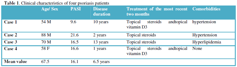
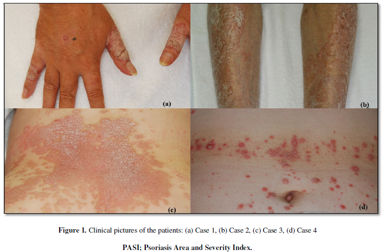
The four patients clinically diagnosed as plaque-type psoriasis
vulgaris were recruited for the investigation (Table 1). Samples of lesional skin were taken using 4 mm punch
biopsy. To confirm the diagnosis of psoriasis, histopathological examination
was performed in all lesional skin samples. Psoriasis Area and Severity Index
(PASI) was determined subjectively by dermatologists. Clinical characteristics
of the patients were obtained from the medical records. Normal skin samples
were obtained from three persons who underwent skin grafting. Informed consent
was given to all participants. Formalin-fixed, paraffin-embedded
tissue samples were cut at 3μm. The
applied antibody was a rabbit polyclonal anti-CCR6 antibody (1:50, clone; LSBio,
WA). Immunohistochemical stains were performed on an automated slide
stainer for immunohistochemistry (Leica BOND-III, Leica Biosystems).
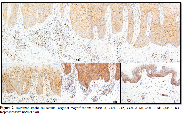
The immunohistochemical results were demonstrated in Figure 2. All psoriatic samples showed
similar results, namely, robust expression of CCR6 in the inflammatory
lymphocytes which had infiltrated into the papillary and perivascular
superficial dermis (Figure 2a-2d).
Although the normal skin contained a few dermal lymphocytes, CCR6 expression in
those cells was less prominent than in the psoriatic samples (Figure 2e). On the other hand, in both
psoriatic and normal skin, keratinocytes in the basal and suprabasal layer of
the epidermis showed comparable CCR6 expression.
There have been a large number of studies
investigating pathophysiological roles of IL-23/ Th17 axis. To our knowledge, only
a few studies of immunohistochemical CCR6 expression in psoriasis patients has been published. Antiga et al.
[2] reported that CCR6-positive and CD4-positive cells were both mainly located
in the superficial dermis, an observation consistent with our own. Moreover, in
our study, immunohistochemical results demonstrated that CCR6 was also
expressed on basal and suprabasal layer of epidermis, which was consistent with
the previous report by Charbonnier et al. [3]. They theorized that
epidermis-expressed CCR6 had a role for a homing of Langerhans cells and a
keratinocyte homeostasis. Although we could not elucidate precise
characteristics of the CCR6-positive infiltrative lymphocytes, it may be possible
that such lymphocytes in the lesional psoriatic skin play a role in promotion
of inflammatory cell migration. Besides, recent studies noted that CCR6 is
expressed on almost all Th17 cells (IL-17A- and IL-22-producing CD4+
T cells). Accordingly, we speculate that the CCR6-positive infiltrative
lymphocytes shown in the lesional psoriatic skin contain some Th17 cells.
ACKNOWLEDGEMENT
This work was supported by Tokyo Medical
University Research Grant.
CONFLICT OF INTEREST
None declared.
- Hedrick
MN, Lonsdorf AS, Hwang ST, et al. (2010) CCR6 as a possible therapeutic
target in psoriasis. Expert Opin Ther Targets 14: 911-922.
- Antiga
E, Volpi W, Chiarini C, et al. (2010) The role of etanercept on the
expression of markers of T helper 17 cells and their precursors in skin
lesions of patients with psoriasis vulgaris. Int J Immunopathol Pharmacol
23: 767-774.
- Charbonnier
AS, Kohrgruber N, Kriehuber E, et al. Macrophage inflammatory protein
3alpha is involved in the constitutive trafficking of epidermal langerhans
cells. (1999) J Exp Med 190: 1755-1768.
-
 Table 1
QUICK LINKS
- SUBMIT MANUSCRIPT
- RECOMMEND THE JOURNAL
-
SUBSCRIBE FOR ALERTS
RELATED JOURNALS
- International Journal of AIDS (ISSN: 2644-3023)
- Oncology Clinics and Research (ISSN: 2643-055X)
- Journal of Forensic Research and Criminal Investigation (ISSN: 2640-0846)
- Journal of Immunology Research and Therapy (ISSN:2472-727X)
- Ophthalmology Clinics and Research (ISSN:2638-115X)
- Journal of Cardiology and Diagnostics Research (ISSN:2639-4634)
- Stem Cell Research and Therapeutics (ISSN:2474-4646)


