1869
Views & Citations869
Likes & Shares
Conclusion: The low prevalence of uterine inversion in our surroundings, particularly in remote regions, has resulted in limited experience in treating this obstetric emergency. The best outcome occurs when uterine inversion is diagnosed early with timely treatment. Nevertheless, the unpredictability of this condition may lead to hysterectomy.
Keywords: Uterine inversion, Atony, Manual uterine re-position, Shoulder dystocia
CASE REPORT
On September 12, 2024, at 03:00 h, a 21-year-old female prime gravida at 42 weeks gestation was admitted to Abdulla Mzee Hospital, complaining of labor pains that had persisted for 5 h. She had good fetal movements and had antenatal profile testing at each of her five prenatal visits. Her blood group was O- Positive, her prenatal hemoglobin level was 10.8%, and HIV and VDRL tests came back negative. She didn't have any noteworthy prior medical or surgical experiences.
Upon assessment, her general condition was fair, not pale, with a BMI of 24.7kg/m2. Fundal height was a term, longitudinal lie, and cephalic presentation. The fetal heart rate appeared to be normal. On vaginal examination, she had a cervical dilatation of 2cm and was admitted to the antenatal ward for observation. On the morning of September 13th, a second evaluation was performed, with a cervical dilatation of 4cm, and all other parameters were normal.A partograph was initiated, and after 8 h of active labor, she experienced an atypical delivery compounded by a delayed second stage of labor due to shoulder dystocia. This necessitates a mediolateral episiotomy and the early use of the shoulder dystocia maneuver. Under teamwork, a male neonate weighing 3100g was successfully delivered with an Apgar score of 9 in the first minute and 10 in the fifth minute. Five minutes during placenta delivery by managing controlled cord traction, the placenta delivered from fundal placentation and passed through the introits.
Uterine inversion was diagnosed because the placenta and membranes were covering a solid mass that was determined to be the uterine cavity. The patient was bleeding heavily and not responding at that time, along with active vaginal bleeding, the amount of blood lost was not measured. Physical examination results in cold extremities, a prolonged capillary refill, tachycardia of 138 beats per minute, tachypnea of 27 breaths per minute, and hypotension of 78/56 mmHg. Examining the genitalia demonstrated complete uterine inversion along with lacerations around the episiotomy incision. Blood work at that time was performed which revealed anemia (hemoglobin 7g/dL, hematocrit 17.4%, erythrocyte 2.07 million/uL) and leukocytosis (19000/uL). Normal thrombocyte level (358.000/uL) was found.
The patient received fluid resuscitation using colloids and crystalloids, as well as a blood transfusion with discontinuation of the uterotonics drug. We successfully performed manual uterine repositioning under general anesthesia in the operating room, followed by 15 min of internal bimanual compression, however, no excessive hemorrhage was detected after repositioning. Therefore, balloon tamponade was not applied. The patient stabilized, and no surgical intervention was required. After the bleeding had been controlled, an episiotomy was repaired.
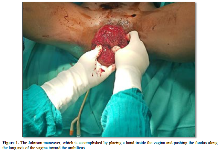
Repeated blood workup following the transfusion of 4 packed red cells shows hemoglobin of 9.8 g/dL, hematocrit of 28%, erythrocyte 3.66 million/uL, leucocyte 16.900/uL, and thrombocyte 150.000/uL. The patient's hemodynamics also stabilized and she regained consciousness. Seven days after admission, the patient recovered without difficulties. She was discharged in stable condition with a hemoglobin level of 10 g/dl and informed of the warning indications and the potential need to return to the hospital in the event of reinvasion, vaginal bleeding, or fever (Figure 1).

DISCUSSION
Acute uterine inversion is an uncommon but life-threatening complication of the third stage of labor in which the uterus is partially or turned inside out [3]. It can be classified by degree, with the first degree- The fundus is within the endometrial cavity, second degree - The fundus protrudes through the cervical opening; third degree -The fundus protrudes to or beyond the introitus and the fourth degree - The uterus and vagina are reversed [4].
Acute uterine inversion occurs within 24 h of delivery, subacute occurs more than 24 h but less than 4 weeks after delivery, and chronic occurs more than 1 month after delivery [5]. Ninety-five percent of uterine inversions happen during puerperal; non-puerperal uterine inversions are typically linked to malignancies outside the uterus [6]. As was the situation in our illustration, when uterine inversion occurred within 24 h of delivery and was of the fourth degree.
The precise cause of uterine inversion is still unknown and up for debate, but it can happen naturally or as a result of mismanagement of the third stage of labor with premature traction of the umbilical cord and fundal pressure before placental separation. Fundal placentation, having an abnormal delivery, prolonged labor, and prime para. Additional factors include congenital weakness or uterine malformations; relaxed uterus, lower uterine segment, and cervix; uterine fibroids; placenta accreta, especially at the uterine fundus; and short umbilical cord [6].
The usage of oxytocin and magnesium sulfate during pregnancy has also been suggested by some research as risk factors for uterine inversion, however, they have not yet been confirmed by Science [7]. Above those, our patients had risk factors for prime para, and their protracted second stage of labor because of shoulder dystocia, with fundal placentation. The clinical appearance of uterine inversion varies depending on its severity and timing. Clinically, incomplete uterine inversion can be modest, whereas total inversion is characterized by profuse vaginal bleeding, inability to palpate the fundus abdominally, and maternal hemodynamic instability. Primarily neurogenic shock is also severe due to nerve tension caused by stretching of the infudibulopelvic ligament and pressure on the ovaries when the fundus moves through the cervical ring [4]. The patient presents with abrupt lower abdomen pain that may or may not be accompanied by a bearing down sensation, supporting the primary clinical diagnosis. The uterine fundus may have a palpable depression or it may not exist at all. Upon vaginal examination, it is typically palpable and/or visible as a dark-reddish-blue bleeding mass at the cervix, vagina, or introitus. As we saw in our case, the medical staff performed admirably and contributed to the mother and infant's survival during delivery. Uterine inversion was diagnosed without the need for ultrasonography confirmation, which is helpful in cases where the clinical examination raises doubts but cannot provide a clear diagnosis. However, Ultrasonography images show signs such as target sign and recognizing an endometrial pseudo strip [8]. The major objective of treatment is to restore maternal hemodynamic stability and prevent fatal outcomes by controlling hemorrhage and doing fluid resuscitation. The goal of first care must be to promptly reverse the uterus as we applied in our reported case (Figure 1). The Johnson maneuver, which accomplished by placing a hand inside the vagina and pushing the fundus along the long axis of the vagina toward the umbilicus. The sooner this operation is performed, the less blood loss will occur and the greater the likelihood of resolution; the longer the interval between the inversion and the procedure, the lower the success rate will be. This lower success rate is brought on by the cervix's involution, which creates a stiff ring that makes it challenging to realign the uterus to its natural position [9].
In addition, utero-relaxant medication and oxytocin infusion suspension are examples of therapeutic interventions. Due to their accessibility and regular use, magnesium sulfate, terbutaline, and salbutamol are the most often recommended medications. Magnesium sulfate takes approximately ten minutes to become effective, whereas terbutaline takes about two minutes. Nitroglycerin (50-500 µg) has been shown by certain writers to produce positive results for cervical ring relaxation. Because halothane, isoflurane, desflurane, and sevoflurane are good tocolytics, in general anesthesia these drugs may be administered when tocolytic medicines are unable to produce uterine relaxation. This is particularly useful in patients with hemodynamic instability due to its fewer effects on hemodynamics as we did in our case Furthermore, hydrostatic reduction can be employed to induce reduction by infusing heated fluid into the vagina under pressure. Other studies describe the use of intravaginal balloons to raise pressure on the uterine fundus and force it back to its original location. Some authors have described it as an alternative when manual reduction fails and surgical intervention is not required [10]. The use of an obstetric suction has also been shown to reverse the uterine fundus. It is imperative to carry out a surgical intervention when conservative therapy is unsuccessful. The literature now in publication describes several procedures, the most frequently reported being laparoscopic, Huntington, Halutzim, and Spinelli. [9] Due to the low occurrence of uterine inversion, no cohort studies large enough to determine the success rate of these procedures have been conducted too far. To avoid a recurrence, uterotonic medicines (oxytocin or misoprostol) must be administered following uterine relocation. A broad-spectrum antibiotic prescription is also indicated to avoid endometritis and sepsis [11] as we did in our instance. Bad enough, prompt management of uterine inversion usually mitigates long-term sequelae. It is unknown whether the condition affects future pregnancy prospects, but case reports exist of uncomplicated pregnancies. However, Women who have experienced uterine inversion need to be counseled that they run the risk of recurrence in subsequent pregnancies [12], therefore, care should be paid during labor monitoring for optimal results.
CONCLUSION
Puerperal uterine inversion is a rare but possibly fatal condition after vaginal birth. A potentially fatal complication that can be avoided by actively and carefully managing the third stage of labor and avoiding cord traction before the appearance of placental separation and fundal pressure. Quick treatment with either non-surgical or surgical procedures can prevent maternal death and other problems.
PATIENT’S PERSPECTIVE
The care provided was timely with a full explanation of the diagnosis and prognosis and a follow-up plan explained.
ACKNOWLEDGMENTS
We are humbly grateful for the support and encouragement given by the Obstetrics/gynecology, pediatric, and anesthetic departments at Abdulla Mzee Hospital.
TIMELINE
The patient was consulted in our hospital and management was initiated. The intervention was done, and the patient was admitted for 1 week. Preparation and completion of the case took one month.
AUTHOR’S CONTRIBUTION
Coauthors contributed to the management of the patient and the writing of the case report. All authors read and approved the final manuscript.
FUNDING
The cost of preparing this manuscript was covered by the Authors.
ETHICAL APPROVAL AND CONSENT TO PARTICIPATE
Written informed consent was obtained from the patient for publication of this case report.
CONSENT FOR PUBLICATION
Written informed consent was obtained from the patient for publication of this case and any accompanying images. A copy of the written consent is available for review by the Editor-in-Chief of this journal. A copy of the clearance document is also available for review by the Editor-in-Chief of this journal.
COMPETING INTERESTS
The authors declare that they have no competing interests.
- Baskett TF (2002) Acute uterine inversion: A review of 40 J Obstet Gynecol Can 24(12): 953-956.
- Rudloff U, Joels LA, Marshall N (2004) Inversion of the uterus at cesarean section. Arch Gynecol Obstet 269: 224-226.
- Sasotya RS, Rinaldi A, Achmad ED, Ma’soem AP, Praharsini K, et (2024) Diagnostics and Management Challenges of Nonpuerperal Uterine Inversions-Case Series. Int J Womens Health 16: 1425-1435.
- Coad SL, Dahlgren LS, Hutcheon JA (2017) Risks and consequences of puerperal uterine inversion in the United States, 2004 through 2013. Am J Obstet Gynecol 217(3): 377.e1-377.e6.
- Ali E, Kumar M (2016) Chronic uterine inversion presenting as a painless vaginal mass at 6 months post-partum: A case report. J Clin Diagn Res 10(5): QD07.
- Das PJ (1940) Inversion of the uterus. BJOG Int J Obstet Gynecol 47(5): 525-548.
- Hostetler DR, Bosworth MF (2000) Uterine inversion: A life-threatening obstetric J Am Board Fam Pract 13(2): 120-123.
- Ida A, Ito K, Kubota Y, Nosaka M, Kato H, et al. (2015) Successful reduction of acute puerperal uterine inversion with the use of a Bakri postpartum balloon. Case Rep Obstet Gynecol 2015(1): 424891.
- Wendel PJ, Cox SM (1995) Emergent obstetric management of uterine Obstet Gynecol Clin North Am 22(2): 261-274.
- Gupta P, Sahu R, Huria A (2014) Acute uterine inversion: A simple modification of hydrostatic method of treatment. Ann Med Health Sci Res 4(2): 264-267.
- Lambert KA, Honart AW, Hughes BL, Kuller JA, Dotters-Katz SK (2023) Antibiotic Recommendations After Postpartum Uterine Exploration or Instrumentation. Obstet Gynecol Surv 78(7): 438-444.
- Thakur M, Thakur A (2018) Uterine
QUICK LINKS
- SUBMIT MANUSCRIPT
- RECOMMEND THE JOURNAL
-
SUBSCRIBE FOR ALERTS
RELATED JOURNALS
- Journal of Oral Health and Dentistry (ISSN: 2638-499X)
- Journal of Pathology and Toxicology Research
- Journal of Rheumatology Research (ISSN:2641-6999)
- Archive of Obstetrics Gynecology and Reproductive Medicine (ISSN:2640-2297)
- International Journal of Radiography Imaging & Radiation Therapy (ISSN:2642-0392)
- Advance Research on Endocrinology and Metabolism (ISSN: 2689-8209)
- International Journal of Diabetes (ISSN: 2644-3031)


