International Journal of Bioprocess and Biotechnological Advancements (ISSN:2380-0259)

Research Article
Comparison Between Trace Element Contents in Macro and Micro Follicular Colloid Goiter using Energy Dispersive X-Ray Fluorescent Analysis
5917
Views & Citations4917
Likes & Shares
Background: Colloid nodular goiter (CNG) is the most common disease of the thyroid, even in non-endemic regions, but an etiology of CNG is unclear. It is known that not only iodine (I) but other trace elements (TE) are involved in goitrogenesis. The present study was performed to clarify the preferential accumulation of some TE either in the colloid or in cells of the thyroid gland.
Methods: Seven TE: bromine (Br), cooper (Cu), iron (Fe), I, rubidium (Rb), strontium (Sr), and zinc (Zn) in the thyroid tissues with diagnosed colloid CNG were prospectively evaluated in 16 patients with macro-follicular CNG and 13 patients with micro-follicular CNG. Control group included thyroid tissue samples from 105 healthy individuals. Measurements were performed using 109Cd and 241Am radionuclide-induced energy-dispersive X-ray fluorescent analysis (EDXRF).
Results: It was found that I predominately accumulate in colloid, whereas Rb and Zn in thyroid cells.
Conclusions: There are considerable changes in TE contents in the goitrous transformed tissue of thyroid, which depend on the histology of goiter.
Keywords: Macro and micro follicular colloid nodular goiter of thyroid, Intact thyroid, Trace elements, Energy-dispersive X-ray fluorescent analysis
Abbreviations: CNG: Colloid Nodular Goiter; TE: Trace Elements; EDXRF: Energy-Dispersive X-Ray Fluorescent Analysis; CRM: Certified Reference Material
INTRODUCTION
Colloid nodular goiter (CNG) is the most common disease of the thyroid, even in non-endemic regions [1]. CNG is clinically detected in about 4% of people older than 30 years [1]. CNG is benign lesion; however, during clinical examination, it can mimic malignant tumors. Furthermore, the origination of CNG can indicate the beginning of malignant transformation of the thyroid gland [2].
Up to now, an etiology of CNG is unclear and probably it is multifactorial [3]. There is opinion that CNG occurs when the thyroid is unable to meet the metabolic demands of the body with sufficient hormone production. The thyroid gland compensates by enlarging, which usually overcomes mild deficiencies of thyroid hormones. For over 20th century, there was the dominant hypothesis that CNG is the simple consequence of iodine (I) deficiency, because I is an essential part of thyroid hormones. However, it was found that CNG is a frequent disease even in those countries and regions where the population is never exposed to I shortage [4]. Moreover, it was shown that I excess has severe consequences on human health and associated with the presence of thyroidal disfunctions and autoimmunity, CNG and diffuse goiter, benign and malignant tumors of gland [5-8]. It was also demonstrated that besides I deficiency and excess many other dietary, environmental, and occupational factors are associated with the CNG incidence [9-11]. Among them a disturbance of evolutionary stable input of many trace elements (TE) in human body after industrial revolution plays a significant role in etiology of thyroidal disorders [12].
Besides I involved in thyroid function, other TE has also essential physiological functions such as maintenance and regulation of cell function, gene regulation, activation or inhibition of enzymatic reactions, and regulation of membrane regulation of cell function, gene regulation, activation or inhibition of enzymatic reactions, and regulation of membrane function [13]. Essential or toxic (goitrogenic, mutagenic, carcinogenic) properties of TE depend on tissue-specific need or tolerance, respectively [13]. Excessive accumulation or an imbalance of the TE may disturb the cell functions and may result in cellular degeneration, death, benign or malignant transformation [13-15].
In our previous studies the complex of in vivo and in vitro nuclear analytical and related methods was developed and used for the investigation of iodine and other TE contents in the normal and pathological thyroid [16-22]. Iodine level in the normal thyroid was investigated in relation to age, gender and some non-thyroidal diseases [23,24]. After that, variations of TE content with age in the thyroid of males and females were studied and age- and gender-dependence of some TE was observed [25-41]. Furthermore, a significant difference between some TE contents in normal and cancerous thyroid was demonstrated [42-47].
Histologically, the CNG is cellular hyperplasia of the thyroid acini. There are two histological types of CNG: macro- and micro-follicular. It is obvious that these two types of CNG have different volume ratios “colloid to cells”.
The present study was performed to clarify the preferential accumulation of some TE either in the colloid or in cells of the thyroid gland. Having this in mind, our aim was to assess the bromine (Br), copper (Cu), iron (Fe), I, rubidium (Rb), strontium (Sr), and zinc (Zn) contents in macro- and micro-follicular CNG tissue using 109Cd and 241Am radionuclide-induced energy-dispersive X-ray fluorescent analysis (EDXRF). A further aim was to compare the levels of these TE in the macro- and micro-follicular CNG separately with those in intact (normal) gland of apparently healthy persons, as well as to find differences between the levels of these TE in the macro- and micro-follicular CNG.
All studies were approved by the Ethical Committees of the Medical Radiological Research Centre (MRRC), Obninsk. All the procedures performed in studies involving human participants were in accordance with the ethical standards of the institutional and/or national research committee and with the 1964 Helsinki declaration and its later amendments, or with comparable ethical standards.
MATERIALS AND METHODS
All patients suffered from СNG (n=29, mean age M±SD was 47±14 years, range 30-64) were hospitalized in the Head and Neck Department of the Medical Radiological Research Centre. Thick-needle puncture biopsy of suspicious nodules of the thyroid was performed for every patient, to permit morphological study of thyroid tissue at these sites and to estimate their TE contents. For all patients the diagnosis has been confirmed by clinical and morphological results obtained during studies of biopsy and resected materials. Histological conclusion for all thyroidal lesions was the macro-follicular CNG (n=16) and micro-follicular CNG (n=13).
Normal thyroids for the control group samples were removed at necropsy from 105 deceased (mean age 44±21 years, range 2-87), who had died suddenly. The majority of deaths were due to trauma. A histological examination in the control group was used to control the age norm conformity, as well as to confirm the absence of micro-nodules and latent cancer.
All tissue samples were divided into two portions using a titanium scalpel [48]. One was used for morphological study while the other was intended for TE analysis. After the samples intended for TE analysis were weighed, they were freeze-dried and homogenized [49]. The pounded sample weighing about 8 mg was applied to the piece of Scotch tape serving as an adhesive fixing backing.
To determine the contents of the TE by comparison with known data for standard, aliquots of commercial, chemically pure compounds and synthetic reference materials were used [50]. The microliter standards were placed on disks made of thin, ash-free filter papers fixed on the Scotch tape pieces and dried in a vacuum. Ten subsamples of the Certified Reference Material (CRM) IAEA H-4 (animal muscle) weighing about 8 mg were analyzed to estimate the precision and accuracy of results. The CRM IAEA H-4 subsamples were prepared in the same way as the samples of dry homogenized thyroid tissue.
Details of the relevant facility for EDXRF with 109Cd radionuclide source for Br, Cu, Fe, Rb, Sr, and Zn determination, and facility for EDXRF with 241Am radionuclide source for I determination, as well as the results of quality control were presented in our earlier publications [21,25,26,51].
All thyroid samples were prepared in duplicate, and mean values of TE contents were used in final calculation. Using Microsoft Office Excel software, a summary of the statistics, including, arithmetic mean, standard deviation, standard error of mean, minimum and maximum values, median, percentiles with 0.025 and 0.975 levels was calculated for TE contents in normal and CNG tissue. The difference in the results between normal thyroid and two groups of CNG (separately macro- and micro-follicular), as well as between two groups of CNG was evaluated by the parametric Student’s t-test and non-parametric Wilcoxon-Mann-Whitney U-test.
RESULTS
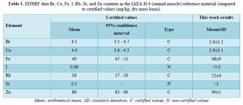
Table 1 depicts our data for seven TE in ten sub-samples of CRM IAEA H-4 (animal muscle) and the certified values of this material.
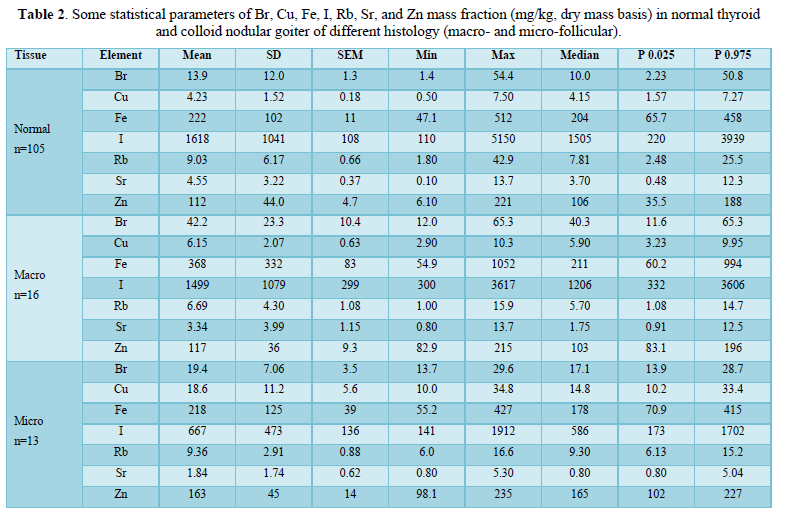
Table 2 presents certain statistical parameters (arithmetic mean, standard deviation, standard error of mean, minimal and maximal values, median, percentiles with 0.025 and 0.975 levels) of the Br, Cu, Fe, Rb, Sr, Zn mass fraction in normal thyroid (n=105), macro-follicular CNG (n=16), and micro-follicular CNG (n=13).
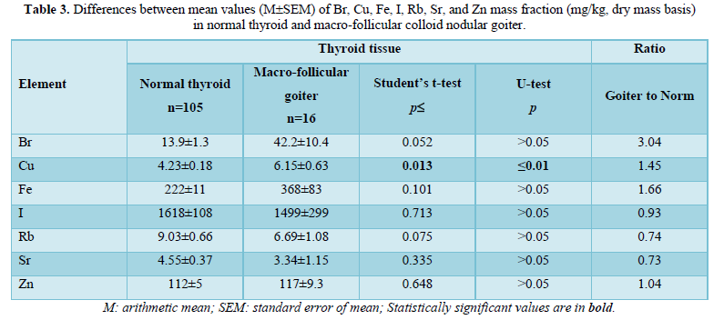
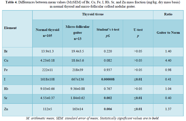
The comparison of Br, Cu, Fe, I, Rb, Sr, and Zn mass fraction in normal thyroid with those in macro- and micro-follicular CNG is shown in Tables 3 & 4, respectively.
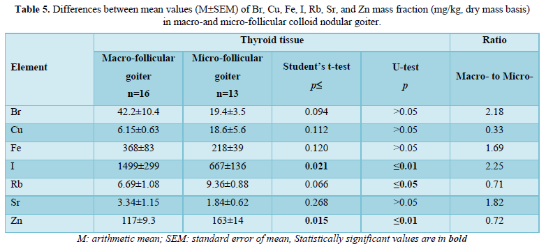
The ratios of means and the difference between mean values of Br, Cu, Fe, I, Rb, Sr, Zn mass fractions in macro- and micro-follicular CNG are presented in Table 5.
DISCUSSION
Precision and accuracy of results
Good agreement of the Br, Cu, Fe, I, Rb, Sr, and Zn contents analyzed by EDXRF with the certified data of CRM IAEA H-4 (Table 1) indicates an acceptable accuracy of the results obtained in the study of TE of the thyroid samples presented in Tables 2-5.
The mean values and all selected statistical parameters were calculated for seven TE (Br, Cu, Fe I, Rb, Sr, and Zn) mass fractions (Table 2). The mass fraction of Br, Cu, Fe, I, Rb, Sr, and Zn were measured in all, or a major portion of normal thyroid and CNG samples.
Effect of goitrous transformation on TE contents
From Table 3, it is observed that in macro-follicular CNG the mass fraction of Cu is 45% higher than in tissues of the normal thyroid. From Table 4, it is observed that in micro-follicular CNG the mass fraction of I and Sr is 59% and 60%, respectively, lower, whereas the mass fraction of Zn is 37% higher than in tissues of the normal thyroid. Thus, if we accept the TE contents in thyroid glands in the control group as a norm, we have to conclude that with a goitrous transformation the Cu, I, Sr, and Zn level in thyroid tissue can be significantly changed.
Association between TE levels and relative volume of colloid and cells
Comparison mass fraction of Br, Cu, Fe, I, Rb, Sr, and Zn in macro- and micro-follicular CNG shown that level of I in macro-follicular goiter is 2.25 times higher, whereas level of Rb and Zn is 29% (only U-test) and 28%, respectively, lower than in micro-follicular goiter. Because the relative volume of colloid in the macro-follicular CNG is higher than in the micro-follicular CNG, it is possible to conclude that I increasingly associated with colloid. On the contrary, because the relative volume of cells in the micro-follicular CNG is higher than in the macro-follicular CNG, it is possible to conclude that Rb and Zn increasingly associated with thyroid cells.
Comparison with published data
The published data on TE contents in the CNG in comparison with normal levels are very scanty and contradictory. For example, Kasanen and Viitanen [52] found elevated levels of Cu in the CNG, but Stojsavljević [9] did not. A significant decrease of the Zn content during goitrous transformation was shown by Błazewicz [53], but in the recent study this change was not confirmed [9]. A relatively good agreement there is only for I, since most of published studies showed the significant decrease of, I content in the CNG [53-56].
Information on the TE contents in macro- or micro-follicular CNG, as well as about the association between TE level and relative volume of colloid and cells in goitrous tissue was not found.
LIMITATIONS
This study has several limitations. Firstly, analytical techniques employed in this study measure only seven TE (Br, Cu, Fe, I, Rb, Sr, and Zn) mass fractions. Future studies should be directed toward using other analytical methods which will extend the list of chemical elements investigated in normal and goitrous thyroid. Secondly, the sample size of macro- or micro-follicular CNG groups was relatively small and prevented investigations of TE contents in CNG group using differentials like gender, stage of disease, and dietary habits of healthy persons and patients with CNG. Lastly, generalization of our results may be limited to Russian population. Despite these limitations, this study provides evidence on goiter-specific tissue Cu, I, Rb, Sr, and Zn level alteration, demonstrates associations between some TE and relative volume of colloid and cells in CNG, and shows the necessity to continue TE research of CNG of different histology.
CONCLUSION
In this work, TE analysis was carried out in the tissue samples of normal and goitrous thyroid using 109Cd and 241Am radionuclide-induced EDXRF. It was shown that 109Cd and 241Am radionuclide-induced EDXRF is an adequate analytical tool for the non-destructive determination of Br, Cu, Fe, I, Rb, Sr, and Zn content in the tissue samples of human thyroid in norm and pathology, including needle-biopsy cores. It was observed the considerable changes in TE contents in the goitrous transformed tissue of thyroid, which depend on the histology of goiter. It was found that I predominately accumulate in colloid, whereas Rb and Zn in thyroid cells.
ACKNOWLEDGEMENTS
The author is extremely grateful to Profs. B.M. Vtyurin and V.S. Medvedev, Medical Radiological Research Center, Obninsk, as well as to Dr. Yu. Choporov, Head of the Forensic Medicine Department of City Hospital, Obninsk, for supplying thyroid samples.
FUNDING
There were no any sources of funding that have supported this work.
CONFLICT OF INTERESTS
The author has not declared any conflict of interests.
- Stuchi LP, Castanhole-Nunes MMU, Maniezzo-Stuchi N, Biselli-Chicote P, Henrique T, et al. (2020) EGFA and NFE2L2 gene expression and regulation by microRNAs in thyroid papillary cancer and colloid goiter. Genes (Basel) 11(9): 954.
- Campbell MJ, Seib CD, Candell L, Gosnell JE, Duh QY, et al. (2015) The underestimated risk of cancer in patients with multinodular goiters after a benign fine needle aspiration. World J. Surg 39: 695-700.
- Frilling A, Liu C, Weber F (2004) Benign multinodular goiter. Scan J Surg 93: 278-281.
- Derwahl M, Studer H (2000) Multinodular goitre: 'much more to it than simply iodine deficiency. Baillieres Best Pract Res Clin Endocrinol Metab 14(4): 577-600.
- Zaichick V (1998) Iodine excess and thyroid cancer. J Trace Elem Exp Med 11(4): 508-509.
- Zaichick V, Iljina T (1998) Dietary iodine supplementation effect on the rat thyroid 131I blastomogenic action. In: Die Bedentung der Mengen- und Spurenelemente. 18. Arbeitstangung. Jena, Friedrich-Schiller-Universität, pp: 294-306.
- Kim S, Kwon YS, Kim JY, Hong KH, Park YK (2019) Association between Iodine Nutrition Status and Thyroid Disease-Related Hormone in Korean Adults: Korean National Health and Nutrition Examination Survey VI (2013-2015). Nutrients 11(11): 2757.
- Vargas-Uricoechea Р, Pinzón-Fernández MV, Bastidas-Sánchez BE, Jojoa-Tobar E, Ramírez-Bejarano LE, et al. (2019) Iodine status in the Colombian population and the impact of universal salt iodization: A double-edged sword? J Nutr Metab 2019: 6239243.
- Stojsavljević A, Rovčanin B, Krstić D, Borković-Mitić S, Paunović I, et al. (2019) Risk assessment of toxic and essential trace metals on the thyroid health at the tissue level: The significance of lead and selenium for colloid goiter disease. Expo Health.
- Fahim YA, Sharaf NE, Hasani IW, Ragab EA, Abdelhakim HK (2020) Assessment of thyroid function and oxidative stress state in foundry workers exposed to lead. J Health Pollut 10(27): 200903.
- Liu M, Song J, Jiang Y, Lin Y, Peng J, et al. (2021) A case-control study on the association of mineral elements exposure and thyroid tumor and goiter. Ecotoxicol Environ Saf 208: 111615.
- Zaichick V (2006) Medical elementology as a new scientific discipline. J Radioanal Nucl Chem 269: 303-309.
- Moncayo R, Moncayo H (2017) A post-publication analysis of the idealized upper reference value of 2.5 mIU/L for TSH: Time to support the thyroid axis with magnesium and iron especially in the setting of reproduction medicine. BBA Clin 7: 115-119.
- Beyersmann D, Hartwig A (2008) Carcinogenic metal compounds: recent insight into molecular and cellular mechanisms. Arch Toxicol 82(8): 493-512.
- Martinez-Zamudio R, Ha HC (2011) Environmental epigenetics in metal exposure. Epigenetics 6(7): 820-827.
- Zaĭchik VE, Raibukhin YuS, Melnik AD, Cherkashin VI (1970) Neutron-activation analysis in the study of the behavior of iodine in the organism. Med Radiol (Mosk) 15(1): 33-36.
- Zaĭchik VE, Matveenko EG, Vtiurin BM, Medvedev VS (1982) Intrathyroidal iodine in the diagnosis of thyroid cancer. Vopr Onkol 28(3): 18-24.
- Zaichick V, Tsyb AF, Vtyurin BM (1995) Trace elements and thyroid cancer. Analyst 120(3): 817-821.
- Zaichick V, Choporov YuYa (1996) Determination of the natural level of human intra-thyroid iodine by instrumental neutron activation analysis. J Radioanal Nucl Chem 207(1): 153-161.
- Zaichick V (1998) In vivo and in vitro application of energy-dispersive XRF in clinical investigations: experience and the future. J Trace Elem Exp Med 11(4): 509-510.
- Zaichick V, Zaichick S (1999) Energy-dispersive X-ray fluorescence of iodine in thyroid puncture biopsy specimens. J Trace Microprobe Tech 17(2): 219-232.
- Zaichick V (2000) Relevance of, and potentiality for in vivo intrathyroidal iodine determination. Ann N Y Acad Sci 904: 630-632.
- Zaichick V, Zaichick S (1997) Normal human intrathyroidal iodine. Sci Total Environ 206(1): 39-56.
- Zaichick V (1999) Human intrathyroidal iodine in health and non-thyroidal disease. In: New aspects of trace element research (Eds: M.Abdulla, M.Bost, S.Gamon, P.Arnaud, G.Chazot). London, Smith-Gordon, and Tokyo, Nishimura, pp: 114-119.
- Zaichick V, Zaichick S (2017) Age-related changes of some trace element contents in intact thyroid of females investigated by energy dispersive X-ray fluorescent analysis. Trends Geriatr Healthcare 1(1): 31-38.
- Zaichick V, Zaichick S (2017) Age-related changes of some trace element contents in intact thyroid of males investigated by energy dispersive X-ray fluorescent analysis. MOJ Gerontol Ger 1(5): 00028.
- Zaichick V, Zaichick S (2017) Age-related changes of Br, Ca, Cl, I, K, Mg, Mn, and Na contents in intact thyroid of females investigated by neutron activation analysis. Curr Updates Aging 1: 5.1.
- Zaichick V, Zaichick S (2017) Age-related changes of Br, Ca, Cl, I, K, Mg, Mn, and Na contents in intact thyroid of males investigated by neutron activation analysis. J Aging Age Relat Dis 1(1): 1002.
- Zaichick V, Zaichick S (2017) Age-related changes of Ag, Co, Cr, Fe, Hg, Rb, Sb, Sc, Se, and Zn contents in intact thyroid of females investigated by neutron activation analysis. J Gerontol Geriatr Med 3: 015.
- Zaichick V, Zaichick S (2017) Age-related changes of Ag, Co, Cr, Fe, Hg, Rb, Sb, Sc, Se, and Zn contents in intact thyroid of males investigated by neutron activation analysis. Curr Trends Biomedical Eng Biosci 4(4): 555644.
- Zaichick V, Zaichick S (2018) Effect of age on chemical element contents in female thyroid investigated by some nuclear analytical methods. Micro Med 6(1): 47-61.
- Zaichick V, Zaichick S (2018) Neutron activation and X-ray fluorescent analysis in study of association between age and chemical element contents in thyroid of males. Op Acc J Bio Eng Bio Sci 2(4): 202-212.
- Zaichick V, Zaichick S (2018) Variation with age of chemical element contents in females’ thyroids investigated by neutron activation analysis and inductively coupled plasma atomic emission spectrometry. J Biochem Analyt Stud 3(1): 1-10.
- Zaichick V, Zaichick S (2018) Association between age and twenty chemical element contents in intact thyroid of males. SM Gerontol Geriatr Res 2(1): 1014.
- Zaichick V, Zaichick S (2018) Associations between age and 50 trace element contents and relationships in intact thyroid of males. Aging Clin Exp Res 30(9): 1059-1070.
- Zaichick V, Zaichick S (2018) Possible role of inadequate quantities of intra-thyroidal bromine, rubidium and zinc in the etiology of female subclinical hypothyroidism. EC Gynaecology 7(3): 107-115.
- Zaichick V, Zaichick S (2018) Possible role of inadequate quantities of intra-thyroidal bromine, calcium and magnesium in the etiology of female subclinical hypothyroidism. Int Gyn Women Health 1(3): IGWHC.MS.ID.000113.
- Zaichick V, Zaichick S (2018) Possible role of inadequate quantities of intra-thyroidal cobalt, rubidium and zinc in the etiology of female subclinical hypothyroidism. Women Health Sci J 2(1): 000108.
- Zaichick V, Zaichick S (2018) Association between female subclinical hypothyroidism and inadequate quantities of some intra-thyroidal chemical elements investigated by X-ray fluorescence and neutron activation analysis. Gynaecol Perinatol 2(4): 340-355.
- Zaichick V, Zaichick S (2018) Investigation of association between the high risk of female subclinical hypothyroidism and inadequate quantities of twenty intra-thyroidal chemical elements. Clin Res: Gynecol Obstet 1(1): 1-18.
- Zaichick V, Zaichick S (2018) Investigation of association between the high risk of female subclinical hypothyroidism and inadequate quantities of intra-thyroidal trace elements using neutron activation and inductively coupled plasma mass spectrometry. Acta Scientific Med Sci 2(9): 23-37.
- Zaichick V, Zaichick S (2018) Trace element contents in thyroid cancer investigated by energy dispersive X-ray fluorescent analysis. Am J Cancer Res Rev 2: 5.
- Zaichick V, Zaichick S (2018) Trace element contents in thyroid cancer investigated by instrumental neutron activation analysis. J Oncol Res 2(1): 1-13.
- Zaichick V, Zaichick S (2018) Variation in selected chemical element contents associated with malignant tumors of human thyroid gland. Cancer Studies 2(1): 2.
- Zaichick V, Zaichick S (2018) Twenty chemical element contents in normal and cancerous thyroid. Int J Hematol Blo Dis 3(2): 1-13.
- Zaichick V, Zaichick S (2018) Levels of chemical element contents in thyroid as potential biomarkers for cancer diagnosis (a preliminary study). J Cancer Metastasis Treat 4: 60.
- Zaichick V, Zaichick S (2018) Fifty trace element contents in normal and cancerous thyroid. Acta Scientific Cancer Biology 2(8): 21-38.
- Zaichick V, Zaichick S (1996) Instrumental effect on the contamination of biomedical samples in the course of sampling. J Anal Chem 51(12): 1200-1205.
- Zaichick V, Zaichick S (1997) A search for losses of chemical elements during freeze-drying of biological materials. J Radioanal Nucl Chem 218(2): 249-253.
- Zaichick V (1995) Applications of synthetic reference materials in the medical Radiological Research Centre. Fresenius J Anal Chem 352: 219-223.
- Zaichick S, Zaichick V (2011) The Br, Fe, Rb, Sr, and Zn contents and interrelation in intact and morphologic normal prostate tissue of adult men investigated by energy-dispersive X-ray fluorescent analysis. X-Ray Spectrom 40(6): 464-469.
- Kasanen A, Viitanen J (1956) On the copper content of the thyroid tissue and serum in thyreotoxicosis. Acta Med Scand 153(6): 467-472.
- Błazewicz A, Dolliver W, Sivsammye S, Deol A, Randhawa R, et al. (2010) Determination of cadmium, cobalt, copper, iron, manganese, and zinc in thyroid glands of patients with diagnosed nodular goiter using ion chromatography. J Chromatogr B Analyt Technol Biomed Life Sci 878(1): 34-38.
- Le Blank AD, Bell RL, Johnson PhC (1973) Measurement of 127J concentration in thyroid tissue by X-Ray fluorescence. J Nucl Med 14(11): 816-819.
- Kohler H, Studer H (1981) Biochemische Veranderungen in “warmen” und “kalten” Strumaknoten. Therapiewoche 31(10): 1539-1545.
- Tadros TG, Maisey MN, Ng Tang Fui SC, Turner P (1981) The iodine concentration in benign and malignant thyroid nodules measured by X-ray fluorescence. Br J Radiol 54(643): 626-629.
-
 Table 1
 -
Table 2
-
Table 3
-
Table 4
-
Table 5


