Research Article
Bone Changes in Condyles of Asymptomatic Temporomandibular Joints & Its Correlation with Age, Gender and Occlusal Condition; a Digital Panoramic Study
873
Views & Citations10
Likes & Shares
Introduction: The TMJ undergoes remodeling throughout lifetime. Several studies on bone changes in patients with TMDs are conducted but limited literature exists on prevalence of bone changes in condyles of asymptomatic TMJs. Hence the present study was proposed to evaluate the prevalence of condylar bone changes among healthy adult Indian population with no complaints of TMDs & correlation with age, gender and dental condition.
Methods: The present study was done on sample size of 250 patients consisting of 5 groups according to age: 20-29, 30-39, 40-49, 50-59 and 60-69 years, 50 patients each (25 males and 25 females). Digital OPG images were analyzed using Digora PCT software. Parameters were recorded and analyzed using chi-square test, paired, independent t-tests and 1-way ANOVA analysis.
Results: The angled shape was most frequent morphology among asymptomatic subjects in this study. The difference between age groups in relation to frequency of right condylar morphology was statistically significant (P<0.05). The prevalence of changes in condylar bone changes was more in individuals above 50 years as compared to those below the age of 40 years. The radiographic bone changes are most commonly observed in older age groups. The condylar width and condylar height asymmetry index in different age groups was statistically significant (P<0.05).
Conclusion: The abnormalities in the condylar morphology and its measurements increases with age. They were seen more frequently in patients with loss of teeth. Condylar width asymmetry in left condyles is more prone to TMDs in different age groups and both genders.
Keywords: Bone density, Condylar symmetry, Eichner’s Index, Horizontal Condylar angle, Mandibular condyle, Orthopantamograph
Abbreviations: OPG: Orthopantomography; PR: Panoramic radiographs; PCT: Program Comprehension Tool; SPSS: Statistical Package for the Social Sciences; TMJ: Temporomandibular Joint; TMDs: Temporomandibular joint disorders
INTRODUCTION
Morphological changes in TMJ occurs as a physiologic process that aims to adapt the structure of the TMJ to meet the functional demands. It is based on an interaction between the mechanical forces sustained by the TMJ and the adaptive capacities of the condyle. The appearance of the mandibular condyle varies greatly among different age groups and individuals. These variations have been reported in symptomatic as well as asymptomatic patients.
Digital panoramic radiography is used for TMJ screening when the clinical examination suggests some form of joint pathology [1] and also for determining gross bony changes in the condyle [2]. It has also been shown that there is consistency and accuracy with digital radiographs of the condylar area. Also, digital imaging with the aid of recent software systems helps in assessing various morphological changes and other bone density parameters that are not possible to measure using conventional radiographs.
Hence the present study was undertaken to evaluate the prevalence and association of bone changes and condylar morphology correlation with age, gender and dental condition among healthy adult Indian population with no complaints of TMDs using digital panoramic radiographs.
MATERIALS AND METHODS
Out Patients visiting the Department of Oral Medicine and Radiology of Dr. Syamala Reddy Dental College, Hospital and Research center, Bangalore. A study sample size of 250 patients consisting of 5 groups according to age: 20-29, 30-39, 40-49, 50-59 and 60-69 years, 50 patients each (25 males and 25 females). The following patients were excluded from the study: when panoramic radiograph did not reveal the condylar anatomy clearly, patients with history of condylar fractures and surgery of condyles, those with developmental anomalies affecting the jaw or syndromes of the craniofacial structures, who had undergone orthodontic treatment, joint disorders like rheumatoid arthritis, gout, infective arthritis, history of parafunctional habits like bruxism, patients with limited mouth opening due to oral submucous fibrosis, space infections, or malignancy of the oral cavity.
Informed consent was taken from the patients prior to the study. The patients were clinically examined, to rule out TMDs based on objective and subjective signs presented. Age and sex information were collected. Dentition was clinically examined and classified into 3 classes as A, B, C using Eichner’s index [3] based on missing teeth in premolar and molar regions. Digital extra oral panoramic images were taken with Soredex Cranex Excel Ceph Panoramic Machine with Exposure Parameters: 73 Kvp; 6 mA and the following parameters were analyzed.
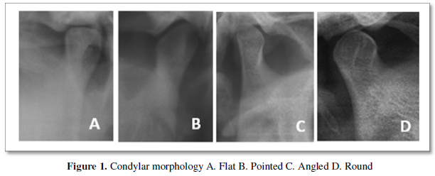
Condylar morphology was analyzed based on the classification of Oliveira-Santos [1] into flat, pointed, angled, round and other shapes (Figure 1).
Condylar bone changes were analyzed according to classification of Uemura [4] as flattening, sclerosis, osteophyte, erosion, deformity, subcortical or ely’s cyst and Surface irregularities (Figure 2).

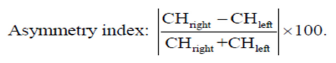
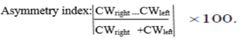
The condylar height asymmetry index and condylar width asymmetry index was measured according to the formula given by Habets [5].
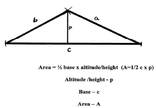
Condylar area (right and left sides separately) was calculated according to the method given by Monalisa [6] in 2007.
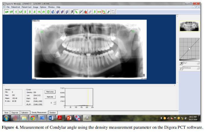
The condylar bone density was measured by marking a square area covering the condyles of both sides and density was measured using the density measurement parameter on the Digora PCT software (Figure 4). For each measure a score of “zero” equated to no demonstrable change,”one” represented mild change, and “two” signified gross change.
RESULTS
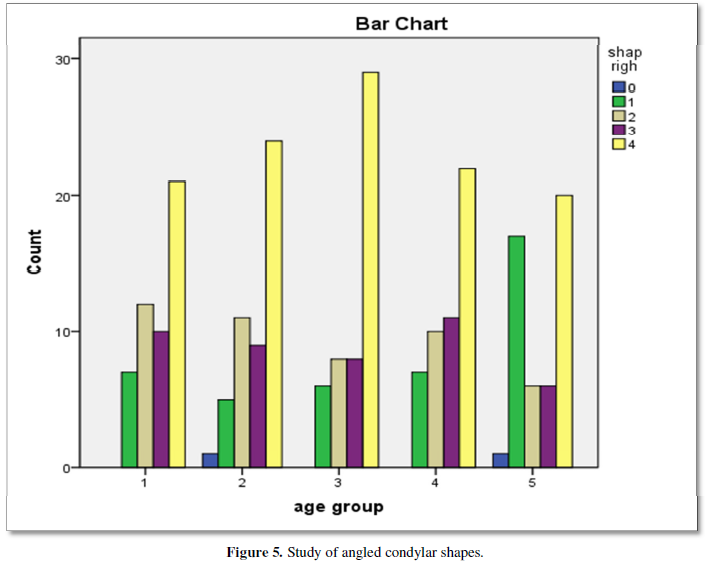
Data were entered into the computer in Microsoft excel sheet 2013 and analyzed using SPSS software version 19. Chi-squared test, paired t test, independent t-test and one-way ANOVA tests were used and p-value less than 0.05, was considered as significant. Overall round and angled condylar shapes were the most frequent among the total number of condyles studied (39.6% and 24.6%, respectively), followed by the pointed shape (18.6%) and flat (17.2%) (Figure 5).
The difference between age-groups in relation to frequency of the morphologic groups was statistically significant (p <0.05). Both genders had round condylar morphology as most common (39.8%), followed by angled condyles (24.4%). The difference between males and females in relation to frequency of the morphologic groups was not statistically significant.
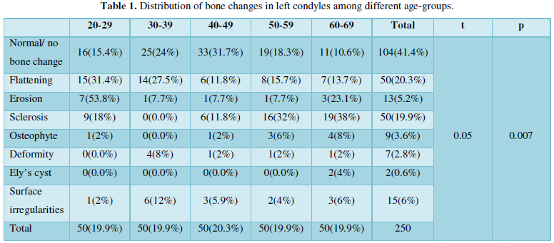
Out of the total 250 patients who were examined radiographically, 130 (52.2%) had radiographic changes in condyles. Sclerosis was observed more in the older age group as compared to the younger age group. When the radiographic bone changes of both condyles together were analyzed with age-groups, statistically significant differences were observed in age group IV and age group V (Table 1).
Out of 250 patients observed for dentition status according to Eichner’s Index, patients with age groups IV (50-59 years old) and age group V (60-69 years), had fewer supporting zones than other age groups. The dentition status among genders showed that both males and females had Class A dentition status with at least 3 to 4 supporting zones. There was no statistically significant difference among these measurements.
The mean condylar width and condylar height asymmetry index in different age groups were statistically significant (p <0.05).
The average condylar density in Indian population was in range of 88 to 91mg/cm3. The observed condylar shapes and radiographic bone changes were significant with condylar height. When comparison was done between dentition status and measurements made on condyles shows a statistically significant difference with condylar height asymmetry index and horizontal angles measured on left condyles.
DISCUSSION
Panoramic radiographs (PR) can be used to determine the condylar changes in asymptomatic TMJs because of low costs, ethical considerations and exposure of patients to relatively low doses of radiation [8].
Overall round and angled condylar shapes were the most frequent among the total number of condyles studied. The results of our study were similar to a study done by Oliveira-Santos [1] in 2009. Despite the absence of subjective symptoms, these variations or abnormalities may act as predisposing factors and may give rise to symptoms following stress, trauma, or iatrogenic insult.
The flat shape was the least frequent morphology among asymptomatic subjects in this study. However, the frequency of flat condyles among symptomatic individuals should be determined in order to suspect any relation to pathological conditions.
It was observed that, as the age increased, the number of condyles affected also increased. This observation is in agreement with the observations of Muir and Goss [9], Huumonen [10] and Takayama [3] that absence of morphologic variation was much more common in the younger age group and age is a factor that determines the degree of remodeling, though there is no direct linear relationship. Also, these findings in right condyles were found to be significant in right condyles, showing that condylar remodeling occurs more in right condyles in healthy adult Indian population.
Various investigators [10-12] have studied the correlation between TMD and both the dental and occlusal relationships. Many authors believe that there is a correlation commonly report that the signs and symptoms of TMD occur readily in individuals who have lost many molars or occlusal supporting areas. Hence the association between changes in the mandibular condyle and dentition status were studied using Eichner’s Index. Of the 250 patients observed for dentition status, patients with age groups IV (50-59 years old) and age group V (60-69 years), had fewer supporting zones than other age groups. Also, there was a significant difference of dentition status with shape, bone changes and horizontal condylar angle in left condyles, showing that left condyles are more prone to TMDs in healthy adult Indian population.
The asymmetry of the mandibular condyle reflects different development of the right and left sides. In 1987-89, Habets [5] proposed in three articles an OPG tracing method, that aimed to investigate any correlation between asymptomatic TMJs and condyle/ramus height and shape. The condylar width and condylar height asymmetry index in different age groups was found to be statistically significant, which was similar to previous studies done by Yáñez-Vico [13] in 2012. Also, high values observed indicating asymmetry in old age groups can be attributed to shape, angular and positional differences between the right and left condyles without any related malocclusion.
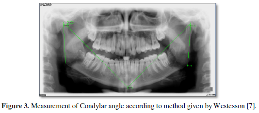
The horizontal angle of inclination of the mandibular condyle is important in maintaining the function of the temporomandibular joint [14]. The horizontal condylar angle was found to be significant among age groups indicating that condylar inclination is more among old age groups, especially in left condyles.
The condylar area was found to be significant among both genders showing that the broader area of the condylar head and glenoid fossa in genders could serve as predictors of future TMDs. The results of our study on condylar area were similar to a study done by Monalisa [6] in 2007.
The condylar density was significant with condylar area showing that the condylar density increases with increase in condylar area. There was a significant difference of dentition status with shape, bone changes and horizontal condylar angle in left condyles, showing that left condyles are more prone to TMDs in healthy adult Indian population.
CONCLUSION
To conclude, abnormalities in the condylar morphology and its measurements increases with age. They were seen more frequently in patients with loss of teeth and left condyles are more prone to TMDs in healthy adult Indian population in different age groups and both genders. Also, condylar width asymmetry among genders could provide basis for the first risk of developing TMDs.
- Oliveira-Santos C, Bernardo RT, Capelozza ALA (2009) Mandibular condyle morphology on panoramic radiographs of asymptomatic temporomandibular joints. Int J Dent 8(3): 114-118.
- Silvestrini-Biavati F, Ugolini A, Laffi N, Canevello C, Silvestrini-Biavati A (2014) Early diagnostic evaluation of mandibular symmetry using orthopantomogram. Indian J Dent Res 25(2): 154-159.
- Takayama Y, Miura E, Yuasa M, Kobayashi K, Hosoi T (2008) Comparison of occlusal condition and prevalence of bone change in the condyle of patients with and without temporomandibular disorders. Oral Surg Oral Med Oral Pathol Oral Radiol Endod 105: 104-112.
- Uemura S, Nakamura M, Iwasaki H, Fuchihata H (1979) A roentgenological study on temporomandibular joint disorders. Morphological changes of TMJ in arthrosis. Dent Radiol 19: 224-237.
- Habets LL, Bezuur JN, Naeiji M, Hansson TL (1988) The Orthopantomogram, an aid in diagnosis of temporomandibular joint problems. II. The vertical symmetry. J Oral Rehabil 15(5): 465-471.
- Monalisa A (2007) An assessment of temporomandibular morphology with routine orthopantomography and the association with temporomandibular disorders. J Syd pp: 1-91.
- Westesson PL, Liedberg J (1987) Horizontal condylar angle in relation to internal derangement of the temporomandibular joint. Oral Surg Oral Med Oral Pathol 64: 391-394.
- Epstein JB, Caldwell J, Black G (2001) The utility of panoramic imaging of the temporomandibular joint in patients with temporomandibular disorders. Oral Surg Oral Med Oral Pathol Oral Radiol Endod 92: 236-239.
- Muir CB, Goss AN (1990) The radiologic morphology of asymptomatic temporomandibular joints. Oral Surg Oral Med Oral Pathol Oral Radiol Endod 70: 349-354.
- Huumonen S, Sipila K, Raustia A M (2007) Panoramic findings in 34-year-old subjects with facial pain and pain-free controls. J Oral Rehab 34(6): 456-462.
- Soikkonen K, Hiltunen K, Ainamo A (1996) Radiographic condylar findings and occlusal imbalance in old people. J Oral Rehab 23(12): 856-859.
- Anuna LM, Amar AS, Keerthilatha MP (2011) Condylar Changes and Its Association with Age, TMD and Dentition Status: A Cross-Sectional Study. Int J Dent 413639: 1-7.
- Yáñez-Vico R-M, Iglesias-Linares A, Torres-Lagares D, Gutiérrez-Pérez J-T, Solano-Reina E (2012) Association between condylar asymmetry and temporo-mandibular disorders using 3D-CT. Med Oral Patol Oral Cir Bucal. 17 (5): e852-e858.
- Larry W, Mandela P, Butt F (2012) Horizontal angle of inclination of the mandibular condyle in a Kenyan population. Anat J Afr 1(1): 46 -49.
-
 Table 1


QUICK LINKS
- SUBMIT MANUSCRIPT
- RECOMMEND THE JOURNAL
-
SUBSCRIBE FOR ALERTS
RELATED JOURNALS
- Journal of Psychiatry and Psychology Research (ISSN:2640-6136)
- Journal of Neurosurgery Imaging and Techniques (ISSN:2473-1943)
- Journal of Cancer Science and Treatment (ISSN:2641-7472)
- Advance Research on Alzheimers and Parkinsons Disease
- International Journal of Diabetes (ISSN: 2644-3031)
- BioMed Research Journal (ISSN:2578-8892)
- Journal of Rheumatology Research (ISSN:2641-6999)


