1134
Views & Citations134
Likes & Shares
Currently there is a
renewed interest in drugs of natural origin simply because they are green
medicine and green medicine offers safe, effective treatment, minimal or no
side effects, easily available, lesser cost and are in great demand in the
developed World health care. There is decreasing in the efficacy of many modern
drugs used for the control of many infections, also an increase in resistance
by several bacteria to various antibiotics and increasing cost of prescribed
drugs. This study aims at investigating the phytochemical constituents and
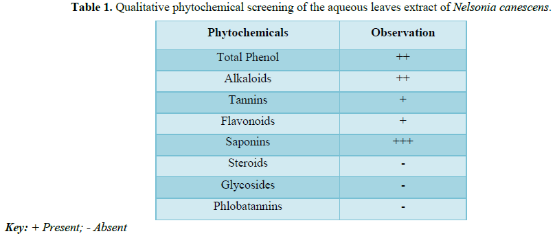
toxicity studies of the aqueous leaves extract of Nelsonia canescens. Results of qualitative phytochemical screening
of the aqueous leaves extract of N.
canescens showed the presence of alkaloids, phenols, tannins, flavonoids
and saponins. The acute toxicity test of the extract N. canescens revealed an oral LD50>2000 mg/kg body weight in
mice. The presence of some of the phytochemicals and the values of the LD50
could explains the plant is being used traditionally for the treatment of a
wide range of illnesses such as fever, pain, chicken pox, measles, constipation
and gastric ulcer without reports of unwanted effects.
INTRODUCTION
Plants are natural reservoir of medicinal
agents almost free from side effects normally caused by synthetic chemicals
[1], medicinal plants play an important role in the health of people living in
rural societies [2], A number of modern drugs have been isolated from natural
sources and many of these isolations were based on the uses of the agents in traditional
medicine [3]. The over use of synthetic drugs with impurities resulting in
higher incidences of adverse drug reactions, has motivated mankind to go back
to nature for safe remedies [4]. The World Health Organization (WHO) estimates
that over 80% of the populations of developing countries currently use
medicinal plants as remedies because of better cultural acceptability, better
compatibility with human body, etc. [5,6]. A number of diseases including
fever, asthma, constipation, esophageal cancer and hypertension have been
treated successfully with herbs [7,8]. In African the use of medicinal plant
has been the unique health care for 4000 years, long before the advent of
western medicine [9]. Currently there is a renewed interest in drugs of natural
origin simply because they are green medicine, and green medicine offer safe,
effective treatment, minimal or no side effect, easily available, lesser cost
and are in great demand in the developed World health care.
Nelsonia canescens (Lam) Spreng. (Family
Acantthaceae) commonly called blue pussy leaf with the synonyms Justicia brunelloides (Lam) [10] is found growing in secondary wet evergreen
forests, savannah forests in open disturbed habitats, especially in moist areas
along road sides, trails, and as a weed in agricultural land [11]. The genus
Nelsonia is classified in the sub-family Nelsonioideae within the Acanthaceae
and has shown to be monophyletic and to comprise the basal lineage among clades
of Acanthaceae [12].
MATERIALS AND METHODS
Sample collection
Animal husbandry
Thirteen
healthy adult Swiss albino mice of both sexes were used for this study. They
were obtained from Niger State Polytechnic Zungeru and were acclimatized for
one (1) week in the Biological garden of School of Science, Federal university
of Science and Technology, Minna Niger state. The mice were housed in
ventilated plastic cages, fed with pellet, deprived of feed overnight before
the administration of test substance and had access to potable water throughout
the period of the study.
Extract preparation
The
fresh leaves of Nelsonia canescens were shade-dried for 21 days at room temperature
(27-29.5°C). The dried leaves were pounded using pestle and mortar into
powdered form at the Centre for Genetic Engineering and Biotechnology (Drug and
Vaccine Discovery Unit), Federal University of Technology Minna, Niger State,
Nigeria. 150 g of the plant powder was soaked in 3 L of distilled water for 72
h and filtered using muslin cloth which was followed by a further filtration
using whatman filter paper No. 1 with pore size of 0.7 µm. The solvent was
removed at 45°C using a rotary evaporator to give a dark solid extract which
weighed 23.7 g. The extract obtained was stored in an air-tight amber bottle
and kept under refrigeration at 4°C prior to further analysis [13].
Phytochemical screening
Phytochemical
screening of the extract of N. canescens
was conducted based on coloration and precipitation tests using standard
methods [14-16].
Test for flavonoids
2
ml of 10% sodium hydroxide was added to 2 ml of the extract in a test tube. A
yellow color which turned colorless upon addition of 2 ml of dilute
hydrochloric acid was an indication a positive result [17].
Test for phenols
2
ml of the extract was mixed with few drops of 10% ferric solution. A greenish
blue or violet or blue coloration was an indication of a positive result [17].
Test for tannins
About
5 drops of 0.1% of ferric chloride (FeCl3) was added to 2 ml of the
extract. A brownish green or blue black coloration was an indication of a
positive result [14].
Test for saponin
2
ml of the extract was diluted with 2 ml distilled water. The mixture was
agitated in a test tube for 4 min. Appearance of about 1 mm layer of foam
indicated a positive result [18].
Test for phelobatannis
2
ml of the extract was boiled with 1% aqueous hydrochloride. Deposition of a red
precipitate indicated a positive result [15].
Test for alkaloids
2
ml of the extract + 2 ml of 10% HCL, to the acidic medium, 2 ml of Meyer’s
reagent were added. Formation of an orange precipitate indicated a positive
result [18].
Test for terpenoids
2
ml of the extract was mixed with 2 ml of chloroform and 1ml of concentrated
sulphuric acid was carefully added to form a layer. A clear upper and lower
with a reddish green inter-phase indicated a positive result [18].
Test for steroids
2
ml of the extract was dissolved in 10 ml of chloroform and then 1 ml of
concentrated sulphuric acid was added by the side of the test tube. Formation
of a reddish upper layer and yellow sulphuric acid layer with green
fluorescence indicated a positive result [18].
Test for anthraquinones
2
ml of the extract was boiled with 5 ml of 10% HCL for 3 min. 5 ml of chloroform
was then added followed by further addition of 5 drops of 10% ammonia. A rose
pink coloration indicates a positive result [16].
Test for glycosides
2
ml of acetic acid, 2 ml of the extract was added. The mixture was cooled in a
cold water bath and then 2 ml of concentrated sulphuric acid was added. Color
development from blue to bluish green indicated a positive result [14].
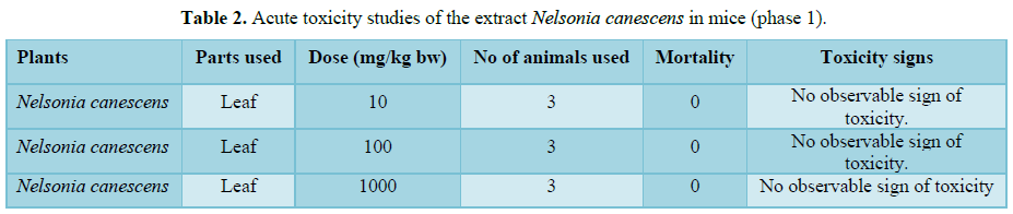
Acute toxicity test
Acute
toxicity test of the plant extract was carried out using the method of Lorke
[19] as described in Latha and Reddy. In the first phase of the experiment, the
animals were randomly divided into three groups of three mice each and were
given the plant extract of 10, 100 and 1000 mg/kg body weight respectively via
oral route. The mice were placed under close observation for 24 h to monitor
all vital signs, behaviors and any mortality before the commencement of the
second phase.
In
phase two, the animals were grouped into three of one animal each and were
orally given higher doses of the plant extract of 1500, 1750 and 2000 mg/kg
body weight. The animals were also observed separately for 24 h for vital
signs, toxicity and mortality.
D0=High
dose that gave no mortality,
D100=Lower
dose that gave no mortality.
RESULTS AND DISCUSSION
This
work is also in agreement with PROTA [24] who reported that aqueous leaf
extract of N. canenscens has
therapeutic and antioxidant properties that the phenolic compounds: tannins
present in the leaf extract of N.
canenscens have the ability to cap the gold nanoparticles by ionic
interaction and thereby stabilizing them [25,26]. Similarly, a number of
researchers have linked the presence of certain phytochemicals in plants as
being responsible for the successful treatment of specific diseases; tannins
and flavonoids are reported to be present in extracts used as antibacterial and
antioxidant [27]. Flavonoids and glycosides are also known to prevent
cardiovascular diseases and ulcers [28]. The presence of alkaloids in many
plant extracts is suggestive of their reasons for a wide range of
pharmacological activities including anti-malaria, antiasthma, anticancer, etc.
[29]. This work is in agreement with other scientific reports that due to the
presence of many phytochemicals in this plant its aqueous extract could be used
for curative activity against many pathogens and therefore explains the use of N. canenscens by many traditionalists in
Africa for the treatment of wide array of illness including malaria.
Results
from Tables 2 and 3 of this study
shows the acute toxicity test of the plant’s aqueous extract of N. canescens is greater than 2000 mg/kg
which means the plant has a very wide safety margin even though the mice were
shivering and became weak at higher doses but no death was recorded even after
24 h. The Organization for Economic Cooperation and Development (OECD) guideline
recommended chemicals labeling and classification of acute toxicity (LD50)
based on oral administration as follows: very toxic substances with an LD50 ≤ 5
mg/kg, while toxic substances having an LD50 to be >5 mg/kg ≤ 50 mg/kg, on
the other hand, harmful substances are said to have an LD50>50 mg/kg ≤ 500
mg/kg and no label >500 mg/kg ≤ 2000 mg/kg, respectively. This work is in
agreement with similar reports of other plants; Sha’a [30] reported that mice
fed with up to 3000 mg/kg body weight of the ethanolic extract of Anacardium
occidentale (cashew) showed sign of weakness but later became active and the
authors concluded that the plant extract is safe. Reports of other
toxicological studies show that dosage up to 5000 mg/kg body weight of
ethanolic extracts of Newbouldia laevis,
aqueous leaf and root extracts of Cymbopogon citrates and crude hydroalcoholic
extracts of Embelia schimperi are
safe [31-33]. However, reported that ethanolic extract of flower of Newbouldia laevis is moderately toxic
because the LD50 in mice was found to be 1264.9 mg/kg body weight when
administered through intra-peritoneal route.
CONCLUSION
In
this present study, the aqueous leafs extract of N. canescens possesses a variety of phytochemical constituents, the
results obtained from acute toxicity studies of the aqueous leafs extract of N. canescens showed that the aqueous
leaves extract of N. canescens did
not produce mortality, signs of toxicity and are relatively safe above 2000
mg/kg body weight. The results of this study suggest that aqueous leafs extract
of N. canescens can be considered a
plant with natural antibiotics at doses used in this work, these scientific
data justifies the traditional use of N.
canescens for treatment of pain, reduce fever, inflammation, constipation
and gastric ulcer. Further studies are recommended for possible identification
of the active ingredients and Isolations of functional group present in the
plant extract.
1.
Fennell CW, Lindsey KL, McGaw LJ, Sparg SG, Stafford
GI, et al. (2004) Assessing African medicinal plants for efficacy and safety:
Pharmacology screening and toxicity. J Ethnopharmacol 94: 205-217.
2.
Focho DA, Ndam WT, Fogne BA (2009) Medicinal plants of
Aguambu-Bamumbu in the Lebialem highlands, southwest province of Cameroon. Afr
J Pharmacol 3: 1-13
3.
Rizvi MMA, Irshad M, Hassadi GE, Younis SB (2009)
Bioefficacies of cassia fistula; An Indian Labrum (Review). Afr J Pharm
Pharmacol 3: 287.
4.
WHO (1999) WHO monographs on selected medicinal plants.
1: 1-295.
5.
Kamboj VP (2000) Herbal medicine. Curr Sci 78: 35-39.
6.
Yadav NP, Dixit VK (2008) Recent approaches in herbal
drug standardization. Int J Intergr Biol 2: 195-203.
7.
Cousins D, Huffman MA (2002) Medicinal properties in
the diet of gorillas: An ethnopharmacolgical evaluation. African Study
Monographs 23: 65-89.
8.
Saganuwan AS (2010) Some medicinal plants of Arabian
peninsula. Afr J Miicrob 4: 766-788.
9.
Silverthorn DU (2010) Fisiologia umana. Un approccio
integrato quinta edizione, pp: 354-358.
10.
Mahias S.N. Ilyas, N. and Musa, K.Y. (2007)
Phytochemical constituents of some medicinal plants use amongst the Takkad
people of Southern Kaduna-Nigeria. Chem Class J CSN Zaria 4: 70-75
11.
McDade LA, Daniel TF, Kiel CA, Borg AJ (2012)
Phylogenetic placement, delimitation and relationships among genera of the
enigmatic Nelsonioideae (Lamiales: Acanthaceae). Taxon 61: 63765.
12.
McDade LA, Daniel TF, Kiel CA (2008) Toward a
comprehensive understanding of phylogenetic relationships among lineages of
Acanthaceae S.L. (Lamiales). Am J Bot 95: 1136-1152.
13.
Mann A (2007) Survey of ethnomedicine for the treatment
of tuberculosis. Chemistry Perspective. Ayanwola Printing.
14.
Sofowora A (1993) Medicinal plant and traditional
medicine in Africa. Lagos-Nigeria: Spectrum books Limited. Standardization of
Herbal Medicines 3: 55-61.
15.
Trease GE, Evans WC (1989) Trease and Evans Mexican
medical plants. J Ethnopharmacol Pharmacogn 13: 222-230.
16.
Harborne JB (1998) Phytochemistry methods. In: Aguide
to modern techniques of plant analysis. 3rd Edn. Champman and Hall
publishing, London. United Kingdom, p: 67.
17.
Trease GE, Evans WC (2002) Pharmacognosy. 15th
Edn. Saunders Publishers, London, pp: 391-393.
18.
Mbatchou VC, Kossono (2012) Aphrodisiac activity of
oils from Anacardium occidentate L.
seeds and seed shells. Phytopharmacology 2: 81-91.
19.
Lorke DA (1983) New approach to practical acute
toxicity testing. Arch Toxicol 54: 275-287.
20.
Doughari JH (2006) Antimicrobial activity of Tamarindus indica Linn. Trop J Pharm
Res, pp: 555-597.
21.
De Krishna MA, Benerjee AB (1999) Antimicrobial
screening of some Indian species. Phytotherapy Res 13: 616-618.
22.
Owoyele VB, Oloriegbe YY, Balogun EA, Soladoye AO
(2005) Analgesic and anti-inflammatory properties of Nelsonia canescens leaf extract. J Ethnopharmacol 99: 153-156.
23.
PROTA (2014) PROTA4U web database. Grubben GJH, Denton
OA, eds. Wageningen, Netherlands: Plant Resources of Tropical Africa. Available
at: http://www.prota4u.org/search.asp
24.
Shittu S (2016) In vitro membranous activity of
biosynthesized gold nanoparticle from aqueous leave extract of Nelsonia canescens. EJMP 15: 1-8.
25.
Singh HP, Gujral SS, Sharma SK, Sharma RK (2015) Tannic
acid: A natural source to tailor nanocrystalline silver particles of different
morphologies as antibacterial agent. Adv Mater Lett 62: 1043-1049.
26.
Cook MC (1996) Flavonoids - Chemistry, metabolism,
cardioprotective effects and dietary sources. Nutr Biochem 7: 66-76.
27.
Swigio G, Tyrakwoska B (2003) Quality of commercial
apple juices evaluated on the basis of polyphenol content and TEAC antioxidant
activity. J Food Sci 68: 1844-1849.
28.
Kittakoop P, Mihidol C, Ruchirawat S (2014) Alkaloids
as important scaffolds in therapeutic drugs for the treatments of cancer,
tuberculosis and smoking cessation. Curr Top Med Chem 14: 239-255.
29.
Sha’a KK, Ajayi OO, Arong GA (2014) The in vitro
antimalarial activity of aqueous and ethanolic extracts of Anacardium occidentale against Plasmodium
falciparum in Damboa, North-Eastern Nigeria. Int J Sci Technol 4: 80-85.
30.
Debebe Y, Tefera M, Mekonnen W, Abebe D, Woldekidan S,
et al. (2015) Evaluation of anthelmintic potential of the Ethiopian medicinal
plant Embelia schimperi vatke in vivo and in vitro against some intestinal parasites. BMC Complement Altern
Med 15: 187.
31.
Arome D, Chinedu E, Ameh SF, Sunday AI (2016)
Comparative anti-plasmodial evaluation of Cymbopogon
citratus extracts in Plasmodium
berghei infected mice. J Curr Res Sci Med 2: 29-35.
32.
Usman H, Yaro AH, Garba MM (2008) Phytochemical and
anti-convulant screening of the ethanolic flower extracts of Newbouldia laevis (Bignoniaceae) in
mice. J Pharmacol Toxicol 3: 127-133.
33.
Adekun O (1978) Atlas of Federal Republic of Nigeria,
Ibadan, Onibonoje pres and Book industries Ltd., p: 12.
-
 Table 1
 -
Table 2
-
Table 3

QUICK LINKS
- SUBMIT MANUSCRIPT
- RECOMMEND THE JOURNAL
-
SUBSCRIBE FOR ALERTS
RELATED JOURNALS
- Advances in Nanomedicine and Nanotechnology Research (ISSN: 2688-5476)
- Journal of Agriculture and Forest Meteorology Research (ISSN:2642-0449)
- Journal of Microbiology and Microbial Infections (ISSN: 2689-7660)
- Journal of Astronomy and Space Research
- Food and Nutrition-Current Research (ISSN:2638-1095)
- Journal of Biochemistry and Molecular Medicine (ISSN:2641-6948)
- Journal of Genomic Medicine and Pharmacogenomics (ISSN:2474-4670)


