2526
Views & Citations1526
Likes & Shares
Nanoparticles
(NPs) are one of the most important tools in the emerging area of nanomedicine.
The behavior of NPs in relevant biological environments, as preclinical setting, may be quite complex due to both their
interactions with biological fluids and the formation of a protein layer,
called protein corona (PC). PC remarkably affects the physicochemical
properties of NPs (size, shape, surface chemistry, aggregation state, etc.) and
consequently their biological fate including their pharmacokinetics,
biodistribution, toxicity and therapeutic efficacy. Strong efforts were applied
to correlate PC composition and observed effect after in vitro/in vivo
experiments, but unfortunately poor reproducibility of data is often assessed.
Biological, chemical and physical properties of NPs along with proteins
composition of PC and features of pathological environments hardly complicate
the study of interactions between PC-NP complexes and their interaction with
target cells. In this contest, in order to
quantitatively and qualitatively evaluate PC adsorbed onto NPs, the optimization of purification/separation
procedures and rigorous and standardized analytical methods become a priority request
to further design tailored nanomaterial able to interact with proteins and
cells in a tunable manner.
Keywords: Nanocarriers,
Molecular size, Nanoparticles, Pharmacokinetics, NP complexes
INTRODUCTION
Nanomedicine is
one of the most active research areas of nanotechnology involving the
application of nanocarriers for the medical prevention, diagnosis, and
treatment of diseases [1]. Polymeric or inorganic nanoparticles (NPs) have the
capacity to incorporate active substances of various characteristics and
protect them from the inhospitable biological environment [2]. Due to their
nanometrical size, NPs show ability to cross tissue barriers and the cell
membrane, thus allowing interaction with the smaller components such as
cellular proteins and other macromolecules [3]. The production of PEG-decorated
NPs could reduce the reticuloendothelial system (RES) uptake, accumulation in
liver, spleen or bone marrow, increasing circulation time and limit
non-specific target uptake ultimately leading to a decrease in toxicity [4].
Further functionalization with targeting ligands, which possess the inherent
ability to facilitate selective binding to cell types, can confer “smart”
properties to NPs [5].
Generally, to
rationally design an efficient nanoparticle-based therapeutic tool able to
selectively transfer drugs to the target site, thus minimizing side effects and
increasing therapeutic compliance, the combination of the nanocarriers
formulative aspects along with a fundamental understanding of the molecular
mechanism involved in regulating nanocarrier-biological interactions is highly
required. In fact, immediately after NPs come into contact with
protein-containing media (such as biological fluids), a layer of proteins,
called the protein corona (PC), is formed on the particle surface. This PC
could remarkably alter the original Np molecular identity affecting their
clearance by RES, cellular uptake, biodistribution and also toxicity.
Furthermore, if the NPs are surface-functionalized with selective ligands, the
absorption of proteins could mask the targeting ability, inhibiting their
biological effects. Thus, it is more than evident that the PC plays a key role
in the interaction of particles with cells after systemic administration [6].
The PC has recently been the subject of
extensive studies aiming to investigate it as complex and multiple layer entity
characterized by proteins that exchange dynamically between the surface and the
surrounding environment (soft corona-SC) and proteins more firmly adsorbed on
the surface of the NPs
(hard corona-HC) [7,8]. An unsolved
As evidence, conflicting reports on
cytotoxicity and biological fates were reported even when similar NPs were
tested [9-12]. For example, Ogawara et al. [13] reported that the PC adsorbed
onto polystyrene NPs prolongs their circulation time, while Nagayama et al.
[14] showed the PC on similar polystyrene NPs is responsible of increased
clearance due to rapid recognition by the scavenger receptors and
internalization by Kupffer cells. Examples of conflicting results can be
found for other biological effects as well (e.g. cytotoxicity, targeting
activity, etc.) [15-18].
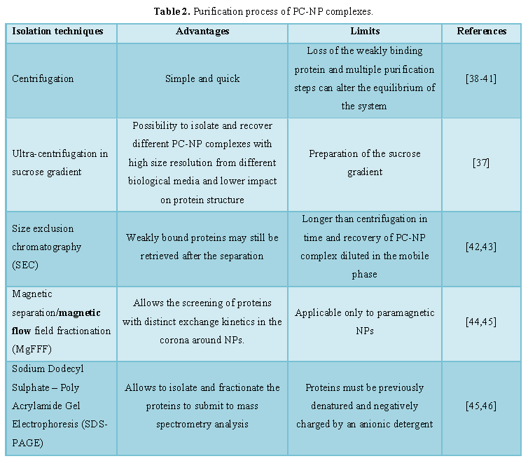
Purification and analyses of
PC-NP complexes
To ameliorate
the difficulty interpreting the biological response observed by using NPs,
researchers focalized the attention optimizing the procedure to characterize
the PC around NPs. In particular, evaluating the overall quantity, density,
thickness composition, relative abundance of each protein, protein binding
affinity, and protein conformation. Firstly, PC-NP complexes were separated
from the protein solution. This operation is not simple as the protein-NPs
interaction is regulated by dynamic exchanges and equilibrium that are particularly
sensitive to purification processes, altering the real contribution of the PC
on the NP surface.
Commonly,
sequential cycles of centrifugation/washing represent the most used method,
because it is simple, suitable and gives reliable results [34,35]. However,
multiple purification steps can alter the equilibrium of complexes and may lead
to modification in corona compositions as previously explain [36]. In
particular, purification techniques adopted to isolate and to study the SC,
which is governed by more dynamic exchanges, must be accurately selected and
optimized in terms of operative conditions (time, temperature, stress, etc.).
Moreover, due to high variability, in order to validate the data, several
replicates must be collected and the results must be statistically relevant.
For this
reason, the development of methodologies that minimize the number of
purification steps of the PC-NP complexes in order to lower the possible impact
of the process on their properties is broadly considered as an urgent issue
[37]. Beyond centrifugation, other techniques, summarized in Table 2, have also been applied to
separate and study PC-NP complexes.
These
techniques were successfully applied to isolate PC-NPs complexes in ex-vivo
experiments, namely by incubating NPs in biological medium simulating relevant
biological conditions. In total, only a few experiments reported reproducible
results in terms of identification of PC isolated from plasma after in vivo administration. Actually, the
idea is to exploit NP features and to adapt or combine different methodologies
in function of the type of NPs. As an example, Sakulkhu et al. [45]
demonstrated that the in vitro PC
profile of polyvinyl alcohol-coated SPIONs differs from in vivo ones, after in vivo
administration, by separating the NPs using a strong external magnetic field
and therefore exploiting the unique magnetic properties of the particles. As
another example, a combined approach was proposed by Hadjidemetriou et al. [47]
to recover lipid-based NPs from the blood circulation of rodents after
intravenous administration to investigate the in vivo PC formation as
well as its evolution. In this case size exclusion chromatography followed by
membrane ultrafiltration allowed the isolation of the PC-NP complex and to
recover reproducible samples to identify the PC components.
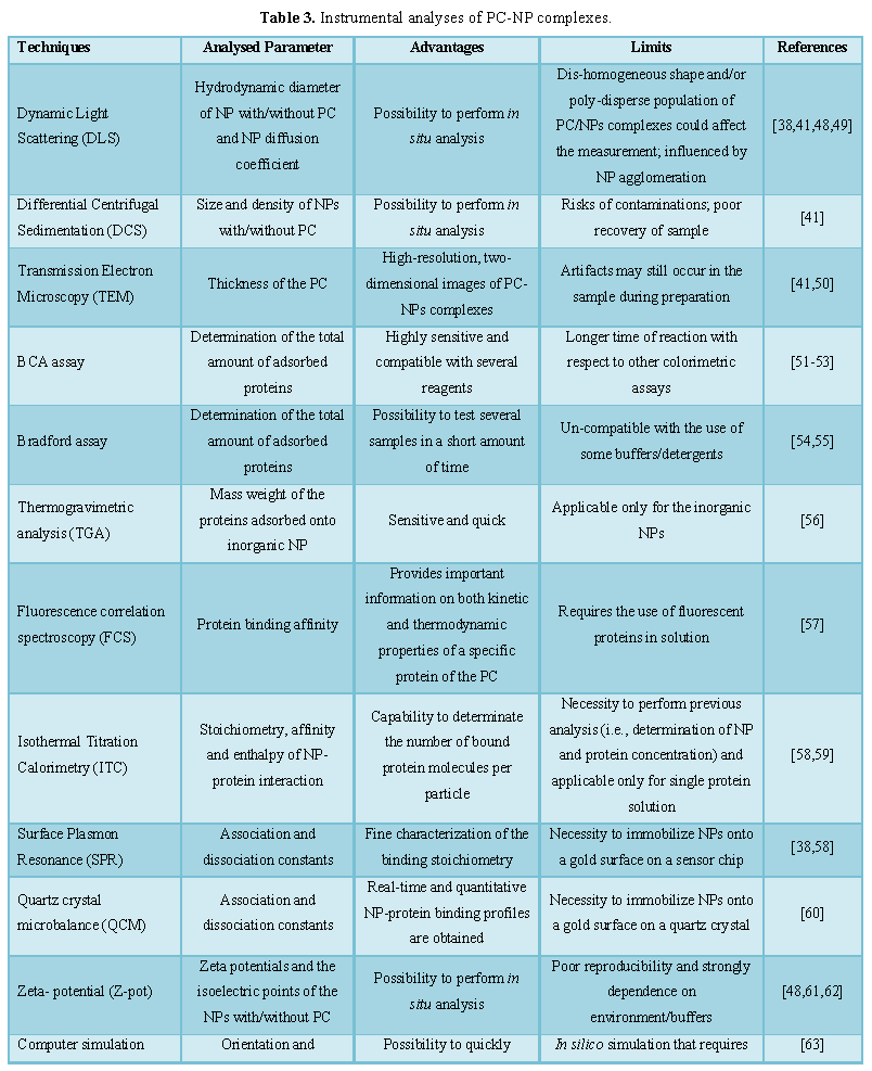
After
purification, several different approaches were proposed to characterize PC-NP
complex in terms size, thickness, quantity, density, thickness composition,
relative abundance of each protein, protein binding affinity and protein
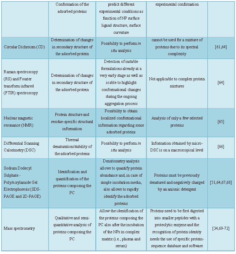
conformation (Table 3).
The interaction between proteins and the NPs
is another relevant issue that could be addressed by different procedures. As
an example, it is possible to consider physical changes in morphology, size and
zeta potential of the complexes with respect to free NPs as proof of a PC-NP
complex presence. Microscopical analyses using transmission electron microscopy
(TEM) and in some cases, atomic force microscopy (AFM), could allow the
visualization of differences in shape, density, surface and also dimensions of
NPs before and after incubation while dynamic light scattering (DLS) rapidly
identifies changes in dimensional distribution and poly-dispersity.
The interaction in terms of affinity,
association and dissociation constant, and the stability of protein adsorbed
were analyzed by using a plenty of techniques including isothermal titration
calorimetry (ITC), circular dichroism, surface plasmon resonance, quartz crystal
microbalance frequently combined together. Gel filtration has been proposed not
only to separate the protein-NP complexes in incubation media as previously
reported, but also to isolate proteins from the NP surfaces as well as to
provide useful information on kinetic exchange rates for adsorbed proteins [58].
Besides proof of the PC-NPs complex presence,
defining the PC represents a major topic: several researchers reported the use
of electrophoresis as the preferred technology, while protein quantity determination was proposed to
be characterized by mass spectrometry, in which protein samples were digested
into small peptides and simply injected into the analysis instrument [73].
In summary, as
evident, the identification and characterization of the PC cannot be obtained
by a single analytical protocol, but different and complementary technologies
are generally combined. In order to reach the highest level of quality and
standardization in PC identification and characterization, the key point relies
on the choice of the techniques in function of both the type of nanocarriers
and on the analytical parameter to investigate. Using multiple characterization
techniques is therefore crucial to analyze different aspects of the PC (i.e.,
presence of complex, composition of PC, reliability in in vivo conditions, etc.) and to get a better understanding of this
biological entity. Remarkably, some techniques allow to detect the protein
corona in situ (ITC),
while other procedures require the detachment of bound proteins from the
nanocarriers before measurements, thus still representing a controversial issue
on PC analysis, since the adoption of purification methods may change
equilibrium properties of the PC.
CONCLUSION
In recent
years, the advance of nanomedicine as applied science in disease treatments
highlighted a deeper need in understanding the interactions between
nanocarriers and the biological environment aiming to improve their
effectiveness and safety profiles. In this context, the study of the PC
connected to any kind of NP drug carrier and its impact on both biodistribution
and interaction with the target site is of extreme importance. Relevant
information of the PC composition could also be exploited in sample screening
at early research stages. This interest generated a wide number of attempted
experiments, but at a deeper analysis of the results, even if remarkable, the
lack of reproducibility and defined protocols in PC analysis still remain an
urgent issue. In particular, as pointed out in this brief review, those data
often obtained in vitro by simulating
biological environments are affected by a high number of variables and seem not
to be reliable and predictive for in vivo
readouts and therefore their translatability. As also pointed out, numerous
purification and investigation techniques were applied to evaluate and
characterize the PC demonstrating that some technologies could really be useful
in analysing some aspects of PC. Thus, in order to concretely exploit PC-NPs
complexes data, the scientific path in this field will surely pass through an
optimization of the protocols with a rational combination of the different
techniques to finalize a systematic and more reproducible PC study approach.
ACKNOWLEDGEMENT
Supported by MAECI grant (PI Tosi,
Nanomedicine for BBB-crossing in CNS oncologic pathologies) and Emilia Romagna
Region grant (Step-by-step screening: RCUP E56C18002000002).
1. Bamrungsap S, Zhao Z, Chen T, Wang
L, Li C, et al. (2012) Nanotechnology in therapeutics: A focus on nanoparticles
as a drug delivery system. Nanomedicine 7: 1253-1271.
2. Fornaguera C, Garcia-Celma MJ
(2017) Personalized nanomedicine: A revolution at the nanoscale. J Pers Med 7.
3. Wilczewska AZ, Niemirowicz K,
Markiewicz KH, Car H (2012) Nanoparticles as drug delivery systems. Pharmacol
Rep 64: 1020-1037.
4. Knop K, Hoogenboom R, Fischer D,
Schubert US (2010) Poly(ethylene glycol) in drug delivery: Pros and cons as
well as potential alternatives. Angewandte Chemie Int 49: 6288-6308.
5. Friedman AD, Claypool SE, Liu R
(2013) The smart targeting of nanoparticles. Curr Pharm Design 19: 6315-6329.
6. Pederzoli F, Tosi G, Vandelli MA,
Belletti D, Forni F, et al. (2017) Protein corona and nanoparticles: How can we
investigate on? Wiley Interdiscip Rev Nanomed Nanobiotechnol 9: e1467.
7. Milani S, Bombelli FB, Pitek AS,
Dawson KA, RäDler J (2012) Reversible versus irreversible binding of
transferrin to polystyrene nanoparticles: Soft and hard corona. ACS Nano 6:
2532-2541.
8. Monopoli MP, Aberg C, Salvati A,
Dawson KA (2012) Biomolecular coronas provide the biological identity of
nanosized materials. Nat Nanotechnol 7: 779-786.
9. Sharifi S, Behzadi S, Laurent S,
Forrest ML, Stroeve P, et al. (2012) Toxicity of nanomaterials. Chem Soc Rev
41: 2323-2343.
10. Mahmoudi M, Lynch I, Ejtehadi MR,
Monopoli MP, Bombelli FB, et al. (2011) Protein-nanoparticle interactions:
Opportunities and challenges. Chem Rev 111: 5610-5637.
11. Mao HY, Laurent S, Chen W, Akhavan
O, Imani M, et al. (2013) Graphene: Promises, facts, opportunities and
challenges in nanomedicine. Chem Rev 113: 3407-3424.
12. Hajipour MJ, Raheb J, Akhavan O,
Arjmand S, Mashinchian O, et al. (2015) Personalized disease-specific protein
corona influences the therapeutic impact of graphene oxide. Nanoscale 7:
8978-8994.
13. Ogawara K, Furumoto K, Nagayama S,
Minato K, Higaki K, et al. (2004) Pre-coating with serum albumin reduces
receptor-mediated hepatic disposition of polystyrene nanosphere: Implications
for rational design of nanoparticles. J Control Release 100: 451-455.
13.
14. Nagayama S, Ogawara K, Minato K,
Fukuoka Y, Takakura Y, et al. (2007) Fetuin mediates hepatic uptake of
negatively charged nanoparticles via scavenger receptor. Int J Pharm 329:
192-198.
15. Su G, Jiang H, Xu B, Yu Y, Chen X
(2018) Effects of protein corona on active and passive targeting of cyclic RGD
peptide-functionalized pegylation nanoparticles. Mol Pharm 15: 5019-5030.
16. Dai Q, Yan Y, Ang CS, Kempe K,
Kamphuis MM, et al. (2015) Monoclonal antibody-functionalized multilayered
particles: Targeting cancer cells in the presence of protein coronas. ACS Nano
9: 2876-2885.
17. Sun D, Gong L, Xie J, Gu X, Li Y,
et al. (2018) Toxicity of silicon dioxide nanoparticles with varying sizes on
the cornea and protein corona as a strategy for therapy. Sci Bull 63: 907-916.
18. Deng ZJ, Liang M, Monteiro M, Toth
I, Minchin RF (2011) Nanoparticle-induced unfolding of fibrinogen promotes
Mac-1 receptor activation and inflammation. Nat Nanotechnol 6: 39-44.
19. Yan Y, Gause KT, Kamphuis MMJ, Ang
CS, O'brien-Simpson NM, et al. (2013) Differential roles of the protein corona
in the cellular uptake of nanoporous polymer particles by monocyte and
macrophage cell lines. ACS Nano 7: 10960-10970.
20. Thiele L, Diederichs JE, Reszka R,
Merkle HP, Walter E (2003) Competitive adsorption of serum proteins at
microparticles affects phagocytosis by dendritic cells. Biomaterials 24:
1409-1418.
21. Borgognoni CF, Mormann M, Qu Y,
Schafer M, Langer K, et al. (2015) Reaction of human macrophages on protein
corona covered TiO(2) nanoparticles. Nanomedicine 11: 275-282.
22. Schöttler S, Becker G, Winzen S,
Steinbach T, Mohr K, et al. (2016) Protein adsorption is required for stealth
effect of poly(ethylene glycol)-and poly(phosphoester)-coated nanocarriers. Nat
Nanotechnol 11: 372-377.
23. Pochert A, Vernikouskaya I,
Pascher F, Rasche V, Linden M (2017) Cargo-influences on the biodistribution of
hollow mesoporous silica nanoparticles as studied by quantitative
(19)F-magnetic resonance imaging. J Colloid Interface Sci 488: 1-9.
24. De Paoli SH, Diduch LL, Tegegn TZ,
Orecna M, Strader MB, et al. (2014) The effect of protein corona composition on
the interaction of carbon nanotubes with human blood platelets. Biomaterials
35: 6182-6194.
25. Shi J, Hedberg Y, Lundin M, Odnevall
Wallinder I, Karlsson H, et al. (2012) Hemolytic properties of synthetic nano
and porous silica particles: The effect of surface properties and the
protection by the plasma corona. Acta Biomater 8: 3478-3490.
26. Salvati E, Re F, Sesana S,
Cambianica I, Sancini G, et al. (2013) Liposomes functionalized to overcome the
blood-brain barrier and to target amyloid-beta peptide: The chemical design
affects the permeability across an in vitro model. Int J Nanomed 8: 1749-1758.
27. Varnamkhasti BS, Hosseinzadeh H,
Azhdarzadeh M, Vafaei SY, Esfandyari-Manesh M, et al. (2015) Protein corona
hampers targeting potential of MUC1 aptamer functionalized SN-38 core-shell
nanoparticles. Int J Pharm 494: 430-444.
28. Caracciolo G, Palchetti S,
Digiacomo L, Chiozzi RZZ, Capriotti AL, et al. (2018) Human biomolecular corona
of liposomal doxorubicin: The overlooked factor in anticancer drug delivery.
ACS Appl Mater Interfaces 10: 22951-22962.
29. Catalano F, Accomasso L, Alberto
G, Gallina C, Raimondo S, et al. (2015) Factors ruling the uptake of silica
nanoparticles by mesenchymal stem cells: Agglomeration versus dispersions,
absence versus presence of serum proteins. Small 11: 2919-2928.
30. Lesniak A, Fenaroli F, Monopoli
MP, Aberg C, Dawson KA, et al. (2012) Effects of the presence or absence of a
protein corona on silica nanoparticle uptake and impact on cells. ACS Nano 6:
5845-5857.
31. Palchetti S, Caputo D, Digiacomo
L, Capriotti AL, Coppola R, et al. (2019) Protein corona fingerprints of
liposomes: New opportunities for targeted drug delivery and early detection in
pancreatic cancer. Pharmaceutics 11.
32. Ge C, Du J, Zhao L, Wang L, Liu Y,
et al. (2011) Binding of blood proteins to carbon nanotubes reduces
cytotoxicity. Proc Natl Acad Sci U S A 108: 16968-16973.
33. Behzadi S, Serpooshan V,
Sakhtianchi R, Muller B, Landfester K, et al. (2014) Protein corona change the
drug release profile of nanocarriers: The “overlooked” factor at the
nanobiointerface. Colloids Surf B Biointerfaces 123:143-149.
34. Sempf K, Arrey T, Gelperina S,
Schorge T, Meyer B, et al. (2013) Adsorption of plasma proteins on uncoated
PLGA nanoparticles. Eur J Pharm Biopharm 85: 53-60.
35. Walkey CD, Chan WC (2012)
Understanding and controlling the interaction of nanomaterials with proteins in
a physiological environment. Chem Soc Rev 41: 2780-2799.
36. Carrillo-Carrion C, Carril M,
Parak WJ (2017) Techniques for the experimental investigation of the protein
corona. Curr Opin Biotechnol 46: 106-113.
37. Di Silvio D, Rigby N, Bajka B,
Mayes A, Mackie A, et al. (2015) Technical tip: High-resolution isolation of
nanoparticle-protein corona complexes from physiological fluids. Nanoscale 7:
11980-11990.
38. Casals E, Pfaller T, Duschl A,
Oostingh GJ, Puntes V (2010) Time evolution of the nanoparticle protein corona.
ACS Nano 4: 3623-3632.
39. Lundqvist M, Stigler J, Cedervall
T, Berggard T, Flanagan MB, et al. (2011) The evolution of the protein corona
around nanoparticles: A test study. ACS Nano 5: 7503-7509.
40. Monopoli MP, Pitek AS, Lynch I,
Dawson KA (2013) Formation and characterization of the nanoparticle-protein
corona. Methods Mol Biol 1025: 137-55.
41. Walczyk D, Bombelli FB, Monopoli
MP, Lynch I, Dawson KA (2010) What the cell “sees” in bionanoscience? J Am Chem
Soc 132: 5761-5768.
42. Lynch I, Cedervall T, Lundqvist M,
Cabaleiro-Lago C, Linse S, et al. (2007) The nanoparticle-protein complex as a
biological entity; a complex fluids and surface science challenge for the 21st
century. Adv Colloid Interface Sci 31: 167-174.
43. Cedervall T, Lynch I, Foy M,
Berggard T, Donnelly SC, et al. (2007) Detailed identification of plasma
proteins adsorbed on copolymer nanoparticles. Angew Chem Int Ed Engl 46:
5754-5756.
44. Ashby J, Pan S, Zhong W (2014)
Size and surface functionalization of iron oxide nanoparticles influence the
composition and dynamic nature of their protein corona. ACS Appl Mater
Interfaces 6: 15412-15419.
45. Sakulkhu U, Mahmoudi M, Maurizi L,
Salaklang J, Hofmann H (2014) Protein corona composition of superparamagnetic
iron oxide nanoparticles with various physico-chemical properties and coatings.
Sci Rep 4: 5020.
46. Lundqvist M, Augustsson C, Lilja
M, Lundkvist K, Dahlback B, et al. (2017) The nanoparticle protein corona
formed in human blood or human blood fractions. PLoS One 12.
47. Hadjidemetriou M, Al-Ahmady Z,
Kostarelos K (2016) Time-evolution of in
vivo protein corona onto blood-circulating PEGylated liposomal doxorubicin
(DOXIL) nanoparticles. Nanoscale 8: 6948-6957.
48. Monopoli MP, Walczyk D, Campbell
A, Elia G, Lynch I, et al. (2011) Physical-chemical aspects of protein corona:
Relevance to in vitro and in vivo biological impacts of
nanoparticles. J Am Chem Soc 133: 2525-2534.
49. Maiorano G, Sabella S, Sorce B,
Brunetti V, Malvindi MA, et al. (2010) Effects of cell culture media on the
dynamic formation of protein-nanoparticle complexes and influence on the cellular
response. ACS Nano 4: 7481-7491.
50. Mahmoudi M, Abdelmonem AM, Behzadi
S, Clement JH, Dutz S, et al. (2013) Temperature: The “ignored” factor at the
nanobio interface. ACS Nano 7: 6555-6562.
51. Gessner A, Lieske A, Paulke BR,
Müller RH (2003) Functional groups on polystyrene model nanoparticles:
Influence on protein adsorption. J Biomed Mater Res Part A 65: 319-326.
52. Goppert TM, Muller RH (2005)
Protein adsorption patterns on poloxamer and poloxamine-stabilized solid lipid
nanoparticles (SLN). Eur J Pharm Biopharm 60: 361-72.
53. Sohaebuddin SK, Thevenot PT, Baker
D, Eaton JW, Tang L (2010) Nanomaterial cytotoxicity is composition, size and
cell type dependent. Particle Fibre Toxicol 7: 1-22.
54. Arvizo RR, Giri K, Moyano D, Miranda
OR, Madden B, et al. (2012) Identifying new therapeutic targets via modulation
of protein corona formation by engineered nanoparticles. PLoS One 7.
55. Mbeh DA, Javanbakht T, Tabet L,
Merhi Y, Maghni K, et al. (2015) Protein corona formation on magnetite
nanoparticles: Effects of culture medium composition and its consequences on
super paramagnetic nanoparticle cytotoxicity. J Biomed Nanotechnol 11: 828-840.
56. Clemments AM, Botella P, Landry CC
(2015) Protein adsorption from biofluids on silica nanoparticles: Corona
analysis as a function of particle diameter and porosity. ACS Appl Mater
Interfaces 7: 21682-21689.
57. Nienhaus GU, Maffre P, Nienhaus K
(2013) Studying the protein corona on nanoparticles by FCS. Methods Enzymol
519: 115-137.
58. Cedervall T, Lynch I, Lindman S,
Berggard T, Thulin E, et al. (2007) Understanding the nanoparticle-protein
corona using methods to quantify exchange rates and affinities of proteins for
nanoparticles. Proc Natl Acad Sci U S A 104: 2050-2055.
59. Winzen S, Schoettler S, Baier G,
Rosenauer C, Mailaender V, et al. (2015) Complementary analysis of the hard and
soft protein corona: Sample preparation critically effects corona composition.
Nanoscale 7: 2992-3001.
60. Kaufman ED, Belyea J, Johnson MC,
Nicholson ZM, Ricks JL, et al. (2007) Probing protein adsorption onto mercaptoundecanoic
acid stabilized gold nanoparticles and surfaces by quartz crystal microbalance
and ζ-potential measurements. Langmuir 23: 6053-6062.
61. Fleischer CC, Payne CK (2014)
Nanoparticle-cell interactions: Molecular structure of the protein corona and
cellular outcomes. Acc Chem Res 47: 2651-2659.
62. Yallapu MM, Chauhan N, Othman SF,
Khalilzad-Sharghi V, Ebeling MC, et al. (2015) Implications of protein corona
on physico-chemical and biological properties of magnetic nanoparticles.
Biomaterials 46: 1-12.
63. Ding HM, Ma YQ (2014) Computer
simulation of the role of protein corona in cellular delivery of nanoparticles.
Biomaterials 35: 8703-8710.
64. Mahmoudi M, Shokrgozar MA, Sardari
S, Moghadam MK, Vali H, et al. (2011) Irreversible changes in protein
conformation due to interaction with superparamagnetic iron oxide
nanoparticles. Nanoscale 3: 1127-1138.
65. Lundqvist M, Sethson I, Jonsson BH
(2004) Protein adsorption onto silica nanoparticles: Conformational changes
depend on the particles' curvature and the protein stability. Langmuir 20:
10639-10647.
66. Brandes N, Welzel PB, Werner C,
Kroh LW (2006) Adsorption-induced conformational changes of proteins onto
ceramic particles: Differential scanning calorimetry and FTIR analysis. J
Colloid Interface Sci 299: 56-69.
67. Gessner A, Lieske A, Paulke BR,
Müller R (2002) Influence of surface charge density on protein adsorption on
polymeric nanoparticles: Analysis by two-dimensional electrophoresis. Eur J
Pharm Biopharm 54: 165-170.
68. Göppert TM, Müller RH (2005)
Polysorbate-stabilized solid lipid nanoparticles as colloidal carriers for
intravenous targeting of drugs to the brain: Comparison of plasma protein
adsorption patterns. J Drug Target 13: 179-187.
69. Pozzi D, Caracciolo G, Capriotti
AL, Cavaliere C, La Barbera G, et al. (2015) Surface chemistry and serum type
both determine the nanoparticle-protein corona. J Proteomics 119: 209-217.
70. Tenzer S, Docter D, Rosfa S,
Wlodarski A, Kuharev JR, et al. (2011) Nanoparticle size is a critical
physicochemical determinant of the human blood plasma corona: A comprehensive
quantitative proteomic analysis. ACS Nano 5: 7155-7167.
71. Docter D, Distler U, Storck W,
Kuharev J, Wunsch D, et al. (2014) Quantitative profiling of the protein
coronas that form around nanoparticles. Nat Protoc 9: 2030-2044.
72. Pederzoli F, Tosi G, Genovese F,
Belletti D, Vandelli Ma, et al. (2018) Qualitative and semi quantitative
analysis of the protein coronas associated to different functionalized
nanoparticles. Nanomedicine (Lond) 13: 407-422.
73. Nguyen VH, Lee BJ (2017) Protein
corona: A new approach for nanomedicine design. Int J Nanomed 12: 3137-3151.
-
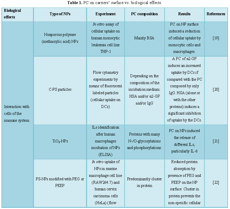
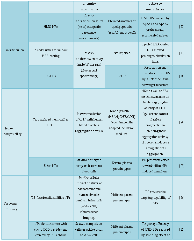
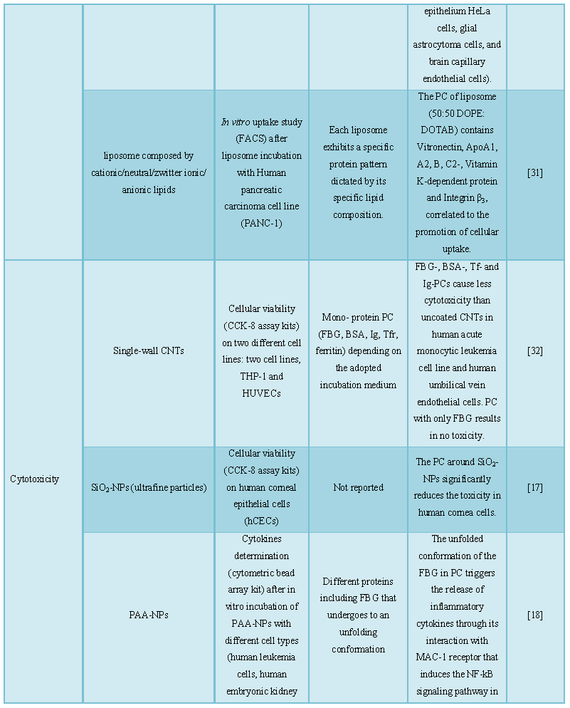
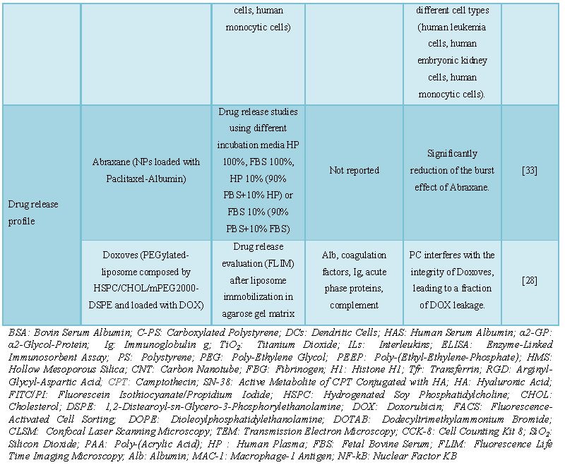
 Table 1
 -
Table 2
-
Table 3
-
Table 4
-
Table 5
-
Table 6
-
Table 7
-
Table 8

QUICK LINKS
- SUBMIT MANUSCRIPT
- RECOMMEND THE JOURNAL
-
SUBSCRIBE FOR ALERTS
RELATED JOURNALS
- Journal of Biochemistry and Molecular Medicine (ISSN:2641-6948)
- Food and Nutrition-Current Research (ISSN:2638-1095)
- Journal of Agriculture and Forest Meteorology Research (ISSN:2642-0449)
- Journal of Microbiology and Microbial Infections (ISSN: 2689-7660)
- Journal of Veterinary and Marine Sciences (ISSN: 2689-7830)
- Journal of Astronomy and Space Research
- Journal of Womens Health and Safety Research (ISSN:2577-1388)


