Review Article
Erythrocyte Aggregation - The Phenomenon of Resting Blood
Katiukhin LN*
Corresponding Author: : Katiukhin LN, Sechenov Institute of Evolutionary Physiology and Biochemistry, Russian Academy of Sciences (IEPhB RAS) 44 Thorez Avenue, Saint-Petersburg, 194223, Russian Federation
Received: October 11, 2018; Revised: December 22, 2018; Accepted: October 18, 2018
Citation: Katiukhin LN. (2018) Erythrocyte Aggregation - The Phenomenon of Resting Blood. J Blood Transfusions Dis, 1(1): 16-19.
Copyrights: ©2018 Katiukhin LN. This is an open-access article distributed under the terms of the Creative Commons Attribution License, which permits unrestricted use, distribution, and reproduction in any medium, provided the original author and source are credited.
Share :
  • 683

    Views & Citations
  • 10

    Likes & Shares

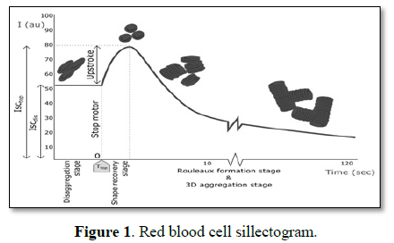
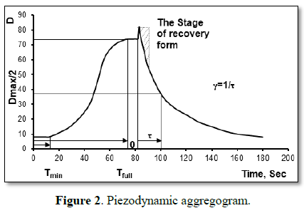
The results of the history of studying the phenomenon of reversible red blood cells aggregation and current methods of measurement have been provided. The work considers the conditions of aggregates formation in vessels with a low shear stress. An assumption was made of inappropriateness of uncontrolled reduction in aggregation capacity of red blood cells for correction of blood flow behavior in case of pathology.


Keywords: Erythrocytes, Aggregability, Shear rate, Aggregometry


Abbreviation: γ: The Rate of Spontaneous Aggregation; τ: The Half-Cycle of Reducing the Photometric Signal; Тmin: Minimum Strength of Aggregates; Тfull: Maximal Strength of Aggregates; ¶Vxy: Speed Derivative (Shear Rate)

HISTORY OF THE PHENOMENON
Under sufficiently low flow rates erythrocytes fasten together along their axis of symmetry and have a tendency to form a continuous structure that is aggregates called rouleaux. The cells adhere with their flat surfaces, which were first found in the records of William Hewson in 1773: “It is necessary to remark, that in a few minutes after particles are spread out on glass, they run in clusters and stick to each other…” [1]. In 1827, other researchers paid attention to this effect in the drop of blood. In 1859, Joseph Lister generalized the observations made and described in detail the results of sound studies under different experimental conditions of this unique property of both red and white blood cells in different kinds of mammals and amphibians. He certified for the first time considerable difference in the property to form strand coins by red blood cells in different kinds of animals, on the one part, red and white blood cells, on the other part, and also strengthening of this process during inflammation [2].

THEORIES OF THE PHENOMENON

It is currently considered that aggregability of red blood cells along with their deformability properties is an important determinant that forms the blood flow properties and structure of the blood. Two parallel models were proposed to explain the aggregation. Under the bridging theory, aggregation is observed when disaggregating forces cannot struggle adsorption of macromolecules by the cells located nearby. The depletion model, based on collinear properties of polymers and experiments in suspension environments with dextrans of different molecular weights, does not put forward any new paradigm as aggregation rises only in presence of dextrans with the weight exceeding 60,000 daltons and is not observed at all in case of lesser weights. Any consideration of the reason for approximation of contacting particles results in depletion of the contact area in large molecules and formation of aggregates, overcoming disruptive forces (electric charge of membranes, mechanic shift, Brownian movement). It should be recognized that formation of red blood cell aggregates seems to result from both osmotic forces and arrangement of cross-links [3-6]. Depletion theory supporters make quite a just conclusion that the properties of red blood cells as internal propensity to aggregation may only be evaluated in artificial conditions that exclude the influence of plasmatic factors, that is, in the solutions of standardized suspending agents or dextrans [7,8]. This effect shows under low shear rates and depends on the properties of red blood cells themselves and physical and chemical properties of suspending agent, that is, on presence   of   macromolecules   in   the   plasma,   such   as fibrinogen, immunoglobulins, etc. [9,10].

METHODS OF MEASUREMENT

In recent years, the flow of literature devoted to the study of this phenomenon has become somewhat weaker. However, there appeared a definite trend of pharmacological companies during development and testing of the drugs intended for improvement of the blood flow properties due to reduction of the erythrocyte aggregation.
Some perfection is made to installations and methods of aggregability characteristics calculation. The aggregation is evaluated with the help of precision installations and adequate methods of foreign and Russian manufacturers. Registration of the so-called sillectograms on RBC aggregometer by Sefam (Brabois-54500 Vandoeuvre les Nancy-France) or modern ektacytometer by Lorrca (LoRRca MaxSis Osmoscan ektacytometer) is done only after rotation stops. Figure 1 presents the relevant sillectogram. The speed of aggregation is found by calculating the peak rise area of extinction after sudden stoppage of Couette cell rotation and shear stress relief during the first 5-10 s. During this period of time, the native RBC form modified in shear stress restored and the stage of the rouleaux formation starts. The maximum shear rate required for complete disaggregation of RBC is calculated [11-13].
The piezo-dynamic method in the microchamber, proposed by the Russian biophysicist, Tukhvatulin from the city of Tomsk, allows giving qualitative characteristics to the total disaggregation-aggregation process. The aggregogram of intact blood, registered on Test photometry device by Optika Design and Technology Institute of RAS Siberian Branch and similar to the one received on Lorrca ektacytometer is presented on Figure 2.
The following indicators are calculated: minimum strength of aggregates Тmin as the time that passed from the moment of launching the forcing influence to the start of movement of separate aggregates and red blood cells in the microchamber; maximum strength Тfull as the time required for complete disaggregation of red blood cells; the rate of spontaneous aggregation (γ) was defined as 1/τ after switching off, where τ is the half-cycle of reducing the photometric signal by half compared to completely disaggregated blood, and, finally, integral aggregation index Tfull [14].

PARTICIPATION OF THE PHENOMENON IN THE FORMATION OF THE RHEOLOGICAL PROPERTIES OF BLOOD

Strengthening of aggregation property of red blood cells in case of inflammation, stress and development of other pathological processes is considered exclusively in the negative context. In particular, it is very popular to note participation of aggregation properties of red blood cells in cases of endothelial disorders, plate’s formation and vessel injuries, when the main events occur near the vessel walls where the shear rate is the highest. Despite the supported interest in evaluation of aggregation properties of red blood cells, let us try to define the appropriateness of such studies [15,16].
The value that determines an internal effort for RBC aggregates destruction is the shear rate, or a shear stress. Multiple observations made since 1966 by Schmid-Schönbein [17] and later other researchers showed that RBC aggregates are destroyed at threshold shear rate approximating the values of 50 s-1. For example, for human vena cava with the diameter of about 10 mm with the least strenuous blood stream conditions, the shear rate is 100 s-1, for veins - 160 s-1, in metabolic vessels, such as venules - (800-1000) s-1, not to speak of arterioles where it reaches 8000 s-1 [15-17].  
The shear rate is calculated as the speed gradient γ=Vx/y in fluid flow tubes in the direction perpendicular to the movement vector Y. Considering parabolic distribution of velocities in the vessel section (Bernoulli effect for viscous fluids flowing slowly in the laminar mode), the shear rate in the vessel clearance is measured linearly, from zero value in the vessel center to the maximum at its wall. Consequently, the true ratio is γthresholdmax=Dthreshold/Dmax. Thus, for vena cava, the ratio of diameters with the threshold shear rate and the maximum reaches 50/100 or ½, that is, Dthreshold=5 mm at Dmax=10 mm. Consequently, the conditions for RBC aggregation phenomenon manifestation in vena cava are only created when the vessel clearance is occluded by 4 times. And this is true to the condition if the blood flow rate will not increase when the clearance narrows. For venules with the diameter of 0.02 mm, this ratio reaches 50/1000, that is, the clearance should decrease by 40 times. For arterioles with the diameter of 0.007 mm, the ratio makes up 50/8000, that is, aggregation is possible if the clearance decreases by more than 25000 times! As for capillaries whose diameter sometimes comes to one third from the RBC diameter and where all the main metabolic processes occur, there is no point in talking about them. It is established that capillaries are never occluded even in experiments with dextrans with large molecular weight [6]. Thus, as aggregates are easily destroyed in the normal blood flow under the influence of slight shear efforts, the tendency for their formation in bloods under physiological conditions in vivo does not considerably influence the blood flow. Thus, the phenomenon of RBC aggregation is shown only in case on complete blood flow stoppage. In our opinion, RBC aggregation is a minor physiological phenomenon comparable with the establishment of hydrogen bonds on the formation of the spatial structure of biological molecules and Van der Waals forces between particles [3,18,19]. Strengthening of aggregation capacity of a large number of oxygen carriers should be considered as the protective reaction of the organism to change in the protein pattern of plasma in case of pathology on the whole and inflammation in particular, which in fact improves blood supply to tissues. Increased aggregation contributes to accumulation of RBC in central areas of vessels, formation of plasmatic lining and sliding layer near endothelial lining. It seems unreasonable to actively fight it. It is appropriate to support the mechanism of struggling, which was generated by the organism in the course of evolution for support of blood flow properties in case of pathological processes. For example, with means of medicines like immunoglobulins, that increases pro-aggregation effect, by concentrating attention on deformation properties of red blood cells. In case of diseases and selection of plasma substitutes, the blood flow properties should be corrected in accordance with the status of a healthy organism, adhering to correlation relationship of RBC flow determinants, i.e., deformability and aggregability for physiological regulation preventing a shift in hemodynamic conditions [12].
On the other hand, there are the evidences that RBCs are important players in hemostasis and thrombosis [20]. In case of vessel injury, destroyed red blood cells help to form a platelet plug and fibrinous clot by activation of RBC coagulation factors (chemical role). They can adsorb and accumulate on their surface plasma coagulation and anticoagulation factors, fibrinolysis, fibrin strands are able fixed to their surface (mechanical role). So, the phenomenon is of theoretical and practical interest as it provides a simple model to study cell interactions in biological systems.
The work was made as part of the state assignment “Physiological and biochemical mechanisms of homeostasis and their evolution”.

1.       Lee K, Kinnunen M, Khokhlova MD, Lyubin EV, Priezzhev AV, et al. (2016) Optical tweezers study of red blood cell aggregation and disaggregation in plasma and protein solutions. J Biomed Opt 21: 35001.

2.       Lister J (1858) On the early stages of inflammation. Phil Trans R Soc Lond 148: 645-702.

3.       Sokolova IA, Yu RS, Shakhnazarov (2011) Erythrocyte aggregation: Some questions and hypotheses. Russian J Biomechanics 1: 7-22.

4.       Chien S, Jan KM (1973) Red cell aggregation by macromolecules: Roles of surface adsorption and electrostatic repulsion. J Supramol Struct 1: 385-409.

5.       Stoltz JF, Donner M (1991) Red blood cell aggregation: measurements and clinical applications. Turk J Med Sci 15: 26-39.

6.       Vicant E (1991) L'agregation erythrocytaire. Sang Thrombose Vaisseaux 6: 377-384.

7.       Baskurt OK, Meiselman HJ (2009) Red blood cell “aggregability”. Clin Hemorheol Microcirc 43: 353-354.

8.       Neu B, Meiselman HJ (2007) Handbook of Hemorheology and Hemodynamics. IOS Press. Amsterdam.

9.       Chien S, Sung LA (1987). Physicochemical basis and clinical implications of red cell aggregation. Clin Hemorheol Microcirc 7: 71-91.

10.    Rampling MW, Meiselman HJ, Neu B, Baskurt OK (2004) Influence of cell‐specific factors on red blood cell aggregation. Biorheology 41: 91-112.

11.    Donner M, Siadat M, Stoltz JF (1988) Erythrocyte aggregation: Approach by light scattering determination. Biorheology 25: 367-376.

12.    Katiukhin LN (2013) Erythrocyte shape transformation in physiological regulation of blood viscosity. Open J Mol Integr Physiol 3: 194-198.

13.    Razavian SM, Del Pino M, Simon A, Levenson J (1992) Increase in erythrocyte disaggregation shear stress in hypertension. Hypertension 20: 247-252.

14.    Tukhvatulin RT, Levtov VA, Shuvaeva VN (1986) Aggregation of erythrocytes in the blood, placed in the macro- and micro-cavity. Sechenov Physiol J 72: 775-785.

15.    Charm SE, Kurland GS (1972) Cardiovascular fluid dynamics. Academic Press, London 2: 15.

16.    Elert G (1998-2017) The Physics Hypertextbook-Viscosity. Physics.info.

17.    Schmid-Schönbein H (1981) Microcirculation: Current physiologic, medical and surgical concepts. Academic Press, N.Y., London, Toronto, Sydney, San Francisco.

18.    Yu SB (1988) Van der Waals forces. Nauka, Moscow, p: 344.

19.    Wong JY, Majewski J, Seitz M, Park CK, Israelachvili JN, et al. (1999). Polymer-cushioned bilayers. I. A structural study of various preparation methods using neutron reflectometry. Biophys J 77: 1445-1447.

20.    Litvinov RI, Weisel JW (2017) Role of red blood cells in haemostssis and thrombosis. ISBT Sci 12: 176-183.