1696
Views & Citations696
Likes & Shares
The aim was to study the effect of the Consciousness
Energy Treated test formulation on vital organ functions viz. bones, heart, liver, lungs, and brain in various cell-based
assays. The test formulation and the cell media were divided into two parts;
one untreated (UT) and other part received the Biofield Energy Treatment
remotely by a renowned Biofield Energy Healer, Alan Joseph Balmer, USA and was
labeled as the Biofield Energy Treated (BT) test formulation/media. Cell
viability data suggested that the test formulation was found as safe and
non-toxic in six different cells. The Biofield Energy Treated medium (BT-Med) +
Biofield Energy Treated Test Item (BT-TI) group showed 181% and 82.2%
restoration of cell viability at 1 and 10 µg/mL, respectively in human cardiac
fibroblasts cells (HCF) compared to the UT-Med + UT-TI group. The UT-Med +
BT-TI group showed 126.8% and 86.3% restoration of cell viability at 10 and 25
µg/mL, respectively with respect to the untreated group in human hepatoma cells
(HepG2). Furthermore, 101.2% (at 10 µg/mL), 103.6% (at 10 µg/mL) and 135% (at
25 µg/mL) restoration of cell viability was observed in adenocarcinomic human
alveolar basal epithelial cells (A549) by UT-Med + BT-TI, BT-Med + UT-TI and
BT-Med + BT-TI groups, respectively compared to the untreated. The alkaline
phosphatase (ALP) level was significantly increased by 90%, 87.3% and 86.9% in
the UT-Med + BT-TI, BT-Med + UT-TI, and BT-Med + BT-TI groups, respectively at
10 µg/mL in human bone osteosarcoma cells (MG-63) compared to the untreated.
Additionally, the level of ALP was significantly increased by 137% in the
BT-Med + UT-TI group in human endometrial adenocarcinoma cells (Ishikawa) at 1
µg/mL compared to the untreated. The percent protection of HCF (heart) cells
(decreased of LDH activity) was significantly increased by 52.1%, 65.9% and
63.5% at 1 µg/mL in the UT-Med + BT-TI, BT-Med + UT-TI, and BT-Med + BT-TI
groups, respectively as compared to the untreated in HCF cells. The percent
protection of HepG2 (liver) cells (decreased of ALT activity) was significantly
increased by 157% and 58.9% at 0.1 and 10 µg/mL, respectively in the BT-Med +
BT-TI group compared to the untreated group in HepG2 cells. The percent
protection of A549 (lungs) cells (increased of SOD activity) was significantly
increased by 168% and 135.4% in the UT-Med + BT-TI group at 10 and 25 µg/mL,
respectively; while, 137% at 10 µg/mL in the BT-Med + UT-TI group as compared
to the untreated group. Serotonin level was significantly increased by 50.8%
(at 63 µg/mL), 78.8% (at 63 µg/mL) and 52.7% (at 1 µg/mL) in the UT-Med +
BT-TI, BT-Med + UT-TI, and BT-Med + BT-TI groups, respectively as compared to
the untreated in human neuroblastoma cells (SH-SY5Y). The relative
quantification (RQ) of vitamin D receptor (VDR) was significantly increased by
265.5% (at 0.1 µg/mL) and 253.4% (at 1 µg/mL) in the UT-Med + BT-TI group;
while 335.3% (at 0.1 µg/mL) in the BT-Med + BT-TI group compared to the
untreated in MG-63 cells. Overall, these results suggest that Biofield Treated
test formulation significantly improved the relevant bones, heart, liver, lungs
and brain-related biomarkers. Altogether data suggest that the Biofield Energy
Treatment (The Trivedi Effect®) can be useful to protect and
maintain the normal function of each vital organ such as lungs, liver, heart,
brain, and bones. Therefore, The Trivedi Effect® can be used as a
complementary and alternative therapy against several disorders such as
coronary artery disease, heart attack, heart failure, arrhythmias, congenital
heart disease, cirrhosis, cardiomyopathy, Wilson disease, liver cancer,
hemochromatosis, pneumonia, asthma, cystic fibrosis, emphysema, chronic
bronchitis, osteoporosis, etc.
INTRODUCTION
Bones, heart, liver,
lungs, and brain disorders are the major concern of human overall health across
the globe. The World Health Organization (WHO) estimates, in 2016, ~17.5
million people die due to cardiovascular (heart) disorders, ~3.5 million people
die due to lungs disorders, ~1.3 million people die due to liver disorders
around the globe each year [1]. Moreover, ~1.2 million people most frequently
diagnosed adult-onset brain disorders in each year in the USA. [2]. Three main
criteria to keep a healthy heart include the opening blood vessels,
strengthening the heart muscle, and controlling free radical damage by
antioxidants [3]. The release of liver mitochondrial enzymes is considered
strong evidence for hepatic (liver) necrosis, which is associated with an
increased production of reactive oxygen species (ROS) that leads to hepatic
lipid peroxidation [4-6]. Oxidative stress in the respiratory system increases
the production of mediators of pulmonary inflammation and initiate or promote
mechanisms of carcinogenesis [7]. The lung is one of the major organs, which is
highly exposed by various oxidants i.e., endogenous and exogenous oxidants
(cigarette smoke, mineral dust, ozone, and radiation). These oxidants produce
free radicals, while reactive oxygen species (ROS) and reactive nitrogen
species (RNS) are produced by phagocytes as well as by alveolar,
polymorphonuclear, bronchial and different endothelial cells [8]. However, the
role of oxidative stress in the pathogenesis of lung diseases has been widely reported
such as asthma, chronic obstructive pulmonary disease (COPD), lung malignancies
and parenchymal lung diseases like idiopathic pulmonary fibrosis and lung
granulomatous diseases [9]. Serotonin (5-hydroxytryptamine, 5-HT) is among the
brain’s neuromodulators responsible for behavior and understanding [10] .Apart
from medicines, non-pharmacologic methods that can increase serotonin by
increasing recognition and happiness and well-being. These factors can protect
against mental and physical disorders [11]. There is currently no universally
accepted test formulation, which improve the organ health biomarkers. With this
respect, the novel test formulation was designed on the basis of best
scientific literature, which is the combination of herbal products viz. Panax
ginseng extract and beta carotene, minerals viz. calcium chloride, magnesium gluconate, zinc chloride, sodium
selenate, ferrous sulfate and vitamins viz.
vitamin B12, vitamin D3, ascorbic acid and vitamin B6. This formulation is
designed for overall functioning of the organs that can result in improved
overall health conditions against many pathological conditions such as lung
disorder, liver disorder, breast cancer, liver cancer, aging, muscle damage,
and overall health. Minerals and vitamins present in the test formulation
provide significant functional support to all the vital organs [12-14]. In
addition, Panax ginseng is one of the
best reported medicinal plants that improve mental, physical abilities,
cognitive health and is potent immunomodulator [15,16].
Various study data
suggested the effect of Energy Therapy in cancer patients through therapeutic
touch [17] massage therapy [18], etc. Complementary and Alternative Medicine
(CAM) therapies are preferred model of treatment, among which Biofield Therapy
(or Healing Modalities) is one approach to enhance emotional, mental, physical,
and human wellness. The National Center of Complementary and Integrative Health
(NCCIH) has recognized and allowed Biofield Energy Healing as a CAM approach in
addition to other therapies and medicines such as natural products,
chiropractic/osteopathic manipulation, Qi Gong, deep breathing, Tai Chi, yoga,
meditation, massage, special diets, healing touch, relaxation techniques,
traditional Chinese herbs and medicines, naturopathy, movement therapy,
homeopathy, progressive relaxation, guided imagery, pilates, acupuncture,
acupressure, Reiki, rolfing structural integration, hypnotherapy, Ayurvedic
medicine, mindfulness, essential oils, aromatherapy, and cranial sacral therapy.
The Human Biofield Energy has subtle energy that has the capacity to work in an
effective manner [19]. CAM therapies have been practiced worldwide with
reported clinical benefits in different health disease profiles [20]. This
energy can be harnessed and transmitted by the practitioners into living and
non-living things via the process of
Biofield Energy Healing. The Biofield Energy Treatment, the Trivedi Effect®,
has been reported to have a significant impact in the field of cancer research
[21,22], materials science [23,24], microbiology [25,26], agriculture [27,28],
nutraceuticals [29,30] and biotechnology [31,32]. Further, the Trivedi Effect®
also significantly improved bioavailability of various low bioavailable
compounds [33-35], an improved overall skin health [36,37], bone health
[38-40], human health and wellness. Based on the excellent outcomes of the
Biofield Energy Therapy in wide spectrum of areas, the authors intend to see
the impact of the Biofield Energy Healing Treated test formulation on the
function of vital organs such as bones, heart, liver, lungs and brain specific
biomarkers in different cell-lines.
METHODS
Chemicals and reagents
Ferrous sulfate,
vitamin B6, vitamin D3, vitamin B12, calcium chloride, naringenin,
trimetazidine (TMZ), 3-(4,5-Dimethylthiazol-2-yl)-2,5-Diphenyltetrazolium
Bromide (MTT) and ethylenediaminetetraacetic acid (EDTA) were obtained from
Sigma Chemical Co. (St. Louis, MO). Zinc chloride, magnesium gluconate,
β-carotene and calcitriol were purchased from TCI chemicals, Japan. Panax ginseng extract obtained from
panacea Phytoextracts, India. Sodium selenate and ascorbic acid were obtained
from Alfa Aesar, India. Silymarin and curcumin were obtained from Sanat
Chemicals, India and quercetin obtained from Clearsynth, India. Reverse Transcription
Kit, RNeasy Mini Kit and Syber Green PCR kits were procured from Quagen, India.
All the other chemicals used in this experiment were analytical grade procured
from India.
Biofield energy healing strategy
The test formulation
was the combination of eleven ingredients viz. calcium chloride, Panax ginseng extract, vitamin B12,
β-carotene, vitamin D3, zinc chloride, magnesium gluconate, sodium selenate,
ferrous sulfate, ascorbic acid and vitamin B6. The test formulation and the
cell media was divided into two parts; one untreated (UT) and other part
received the Biofield Energy Treatment remotely by a renowned Biofield Energy
Healer, Alan Joseph Balmer, USA under laboratory conditions for ~3 minutes
through healer’s unique Biofield Energy Transmission process and was labeled as
the Biofield Energy Treated (BT) test formulation/media. Further, the untreated
group was treated with a “sham” healer for comparison purposes. The “sham”
healer did not have any knowledge about the Biofield Energy Healing Treatment.
The Biofield Energy Healer was located in the USA; however the test items were
located in the research laboratory of Dabur Research Foundation, New Delhi,
India. Biofield Energy Healer in this experiment did not visit the laboratory,
nor had any contact with the test samples. After that, the Biofield Energy
Treated and untreated test items were kept in similar sealed conditions and
used for the study as per the study plan.
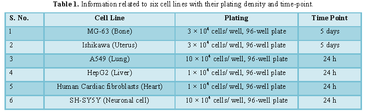
Assessment of cell viability using MTT assay
Cells were counted
using hemocytometer and plated in 96-well plates at the specific density
described in Table 1. The cells were then incubated overnight under growth
conditions to allow cell recovery and exponential growth. Following overnight
incubation, cells were treated with different concentrations of test
formulations (BT/UT). Following respective treatments, cells were incubated in
a CO2 incubator at 37°C, 5% CO2 and 95% humidity and
incubated for time period mentioned in Table
1. After incubation, the plates were taken out and 20 µL of 5 mg/mL of MTT
3-(4,5-dimethythiazol-2-yl)-2,5-diphenyl tetrazolium bromide solution was added
to all the wells followed by additional incubation for 3 h at 37°C. The
supernatant was aspirated and 150 µL of DMSO was added to each well to dissolve
formazan crystals. The absorbance of each well was read at 540 nm using Synergy
HT microplate reader. The percentage cytotoxicity at each tested concentration
of TI was calculated using Equation 1:
%
Cytotoxicity = [(R-X)/R] *100............ (1)
Where, X=Absorbance
of treated cells; R=Absorbance of untreated cells
The concentrations
exhibiting percentage cytotoxicity <30% were considered as non-cytotoxic
[41].
Evaluation of the cytoprotective effect of the
formulation
Cells (human cardiac fibroblasts-HCF; human hepatoma cells-HepG2; and
adenocarcinomic human alveolar basal epithelial cells-A549) were counted and
plated in suitable medium followed by overnight incubation. The cells were then
treated with the test items/positive control at the non-cytotoxic
concentrations for 24 h. After 24 h, oxidative stress was given to the cells
using 10 mM t-BHP for 3.5 h. The untreated cells served as a control
that did not receive any treatment and was maintained in cell growth medium
only. Cells treated with 10 mM of t-BHP alone served as negative
control. After 3.5 h of incubation with t-BHP the above plates were
taken out and cell viability was determined by MTT assay. The percentage
protection corresponding to each treatment was calculated using Equation 2:
% Protection =
[(Absorbancesample-Absorbancet-BHP)]*100/ [Absorbanceuntreated-Absorbancet_BHP]...............
(2)
Assessment of
alkaline phosphatase (ALP) activity
The cells
(human bone osteosarcoma cells-MG-63 and human endometrial adenocarcinoma
cells-Ishikawa) were counted using a hemocytometer and plated in 24-well plates
at the density corresponding to 1 × 10⁴ cells/well in phenol-free
DMEM supplemented with 10% CD-FBS. Following the
respective treatments, the cells in the above plate were incubated for 48 h in CO2 incubator at
37°C, 5% CO2 and 95% humidity.
After 48 h of incubation, the plates were taken out and processed for the
measurement of ALP enzyme activity. The cells were washed with 1x PBS and lysed
by freeze-thaw method, i.e., incubation at -80°C for 20 min followed by
incubation at 37°C for 10 min. To the lysed cells, 50 µL of substrate solution,
i.e., 5 mM of p-nitrophenyl phosphate
(pNPP) in 1 M diethanolamine and 0.24
mM magnesium chloride (MgCl2) solution (pH 10.4) was added to all
the wells followed by incubation for 1 h at 37°C. The absorbance of the above
solution was read at 405 nm using Synergy HT microplate reader (Biotek, USA).
The absorbance values obtained were normalized with substrate blank (pNPP solution alone) absorbance values.
The percentage increase in ALP enzyme activity with respect to the untreated
cells (baseline group) was calculated using Equation 3:
% Increase in ALP =
{(X-R)/R}*100 (3)
Where, X=Absorbance of cells corresponding to positive control and test
groups; R=Absorbance of cells corresponding to baseline group (untreated cells)
Estimation of
lactate dehydrogenase (LDH) in human cardiac fibroblasts (HCF)
The human
cardiac fibroblasts (HCF) Cells were counted and plated at the
density of 0.25 × 10⁶
cells/well in 24-well plates in cardiac fibroblast specific medium followed by overnight incubation.
The cells were then treated with the test formulation/positive control at the
non-cytotoxic concentrations for 24 h. After 24 h, oxidative stress was given
to the cells using 10 mM t-BHP for
3.5 h. The untreated cells were served as control that did not receive any
treatment and were maintained in cell growth medium only. Cells treated with 10
mM of t-BHP alone served as the
negative control. After 3.5 h of incubation with t-BHP the above plates were taken out and LDH activity was
determined using LDH activity kit as per manufacturer’s instructions. The
percent increase in LDH activity was calculated using Equation 4.
%
Increase = [(LDH activitysample-LDH activityt-BHP)]*100/
[LDH activityuntreated-LDH activityt_BHP]..............
(4)
Estimation of ALT in liver cells (HepG2)
The human hepatoma cells (HepG2) were
counted and plated at the density of 5 × 10⁴ cells/well in 48-well plates
in DMEM media followed by overnight incubation. The cells were then
treated with the test formulation/positive control at the non-cytotoxic
concentrations for 24 h. After 24 h, oxidative stress was given to the cells
using 400 µM t-BHP for 3.5 h. The
untreated cells served as control that did not receive any treatment and were
maintained in cell growth medium only. Cells treated with 400 µM of t-BHP alone served as negative control.
After 3.5 h of incubation with t-BHP
the above plates were taken out and ALT activity was determined using ALT
activity kit as per manufacturer’s instructions. The percent increase in ALT
activity was calculated using Equation 5.
%
Increase = [(ALT activitysample-ALT activityt-BHP)]*100/
[ALT activityuntreated-ALT activityt_BHP].............
(5)
Estimation
of superoxide dismutase (SOD) in lung (A549) cells
The adenocarcinomic human alveolar
basal epithelial cells (A549) were counted and plated at the density of 1 × 10⁴
cells/well in 24-well plates in DMEM followed by overnight
incubation.
The cells were then treated with the test formulation/positive control at the
non-cytotoxic concentrations along with 100 µM t-BHP to induce oxidative stress. The untreated cells served as
control that did not receive any treatment and were maintained in cell growth
medium only. Cells treated with 100 µM of t-BHP alone served as negative
control. After 24 h of incubation with t-BHP
the above plates were taken out and SOD activity was determined using SOD
activity kit as per manufacturer’s instructions. The percent increase in SOD
activity was calculated using Equation 6:
%
Increase in SOD activity
= ((X-R)/R)*100................ (6)
Where, X=SOD activity corresponding to test item or positive control;
R=SOD activity corresponding to control group
Estimation
of serotonin in neuronal cells (SH-SY5Y)
The human neuroblastoma (SH-SY5Y)
cells were counted and plated at the density of 10 × 104
cells/well in 96-well plates followed by overnight
incubation.
The cells were then treated with the test items/positive control at the
non-cytotoxic concentrations. The untreated cells served as control that did
not receive any treatment and were maintained in cell growth medium only. The
treated cells were incubated for 24 h. Serotonin release was determined by ELISA
as per manufacturer’s protocol. The percent increase in serotonin levels was
calculated using Equation 7.
[(X-R)/R]*100................ (7)
Where, X=Serotonin levels corresponding to
test item or positive control; R=Serotonin levels corresponding to control
group
Effect of test
formulation on vitamin D receptor (VDR) in bone (MG-63) cells
The human bone osteosarcoma (MG-63)
cells were counted using the hemocytometer were plated at a density of 2
× 105 cells/well in 6-well plates followed by
overnight incubation. The cells were then sera starved for 24 h and treated with the test
formulation/positive control at the non-cytotoxic concentrations. The untreated
cells that served as control that did not receive any treatment and were
maintained in cell growth medium only. The treated cells were incubated for 24
h and VDR expression was determined by
Q-PCR using VDR specific primers. Cells were harvested by scrapping and
washed with PBS. Cell pellets obtained were analyzed for VDR gene expression
using human VDR specific primers: Forward: 5’-GCTGACCTGGTCAGTTACAGCA-3’,
Reverse: 5’-CACGTCACTGACGCGGTACTT-3’. VDR gene expression was normalized using
House-keeping (HK) reference. Relative quantification (RQ) of VDR gene in
Biofield Energy Treated cells was calculated with respect to the untreated
cells using Equation 8:
RQ = 2-N (8)
Where N is the relative Threshold Cycle (CT)
value of treated sample with respect to the untreated sample.
STATISTICAL
ANALYSIS
All the values were represented as Mean ± SD
(standard deviation) of three independent experiments. The statistical analysis
was performed using Sigma Plot statistical software (v11.0). For two groups
comparison Student’s t-test was used.
For multiple group comparison, one-way analysis of variance (ANOVA) was used
followed by post-hoc analysis by Dunnett’s test. Statistically significant
values were set at the level of p ≤
0.05.
Results and Discussion
Cell
viability using MTT assay
Determination of non-cytotoxic concentration
of the test formulation and positive controls by MTT cell viability assay was
used in terms of percent viable cells in six (6) different cell-lines viz. MG-63, Ishikawa, A549, HepG2, HCF
and SH-SY5Y. Based on the percent cell viability data, it was observed that the
formulation and positive controls were safe and non-toxic at the tested
concentrations in six different cell lines and selected for other parameters
analysis.
Evaluation of cytoprotective effect
of the test formulation
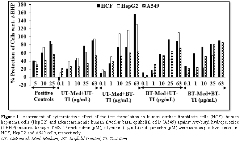
Effect of
the test formulation on vital organs viz.
heart, liver, and lungs using cell-based assay under the stimulation of tert-butyl hydroperoxide (t-BHP) induced oxidative stress. t-BHP has been regularly used for the
induction of oxidative stress in various cells [41]. The cytoprotective
activity of the Biofield Energy Treated test formulation on the restoration of
cell viability was determined against t-BHP
induced cell damage and the result is shown in Figure 1. Trimetazidine (TMZ) was used as positive control in human
cardiac fibroblasts cells (HCF) and showed, restoration of cell viability by
40.57%, 60.68% and 90.04% at 5, 10 and 25 µg/mL, respectively compared to the
t-BHP induced group. Besides, the test formulation showed 60.5% and 181%
restoration of cell viability at 1 µg/mL in the UT-Med + BT-TI and BT-Med +
BT-TI groups, respectively as compared to the UT-Med + UT-TI group. Moreover,
at 10 µg/mL the UT-Med + BT-TI, BT-Med + UT-TI and BT-Med + BT-TI groups showed
80.7%, 38.9% and 82.2% restoration of cell viability, respectively than UT-Med
+ UT-TI group. Additionally, the test formulation showed 20.5% restoration of
cell viability at 25 µg/mL in the BT-Med + UT-TI group as compared to the
UT-Med + UT-TI group. Further, at 63 µg/mL the test formulation showed 71.8%
restoration of cell viability in the UT-Med + BT-TI group than UT-Med + UT-TI
group (Figure 1). Silymarin was used
as positive control in human hepatoma
cells (HepG2) resulted, restoration of cell viability by
38.79%, 73.92% and 81.74% at 5, 10 and 25 µg/mL, respectively compared to the
t-BHP induced group. The test formulation showed 64.4%, 28.5%, 126.8% and 86.3%
restoration of cell viability at 0.1, 1, 10 and 25 µg/mL, respectively in the
UT-Med + BT-TI group as compared to the UT-Med + UT-TI group. Moreover, at 63
µg/mL the UT-Med + BT-TI and BT-Med + UT-TI groups showed 39.3% and 16.6%
restoration of cell viability, respectively than UT-Med + UT-TI group (Figure 1). Quercetin was used as
positive control in adenocarcinomic
human alveolar basal epithelial cells (A549) resulted,
restoration of cell viability by 31.24%, 41.93% and 55.74% at 5, 10 and 25
µg/mL, respectively compared to the t-BHP induced group. Besides, the test
formulation showed 45.4% and 19.5% restoration of cell viability in the UT-Med
+ BT-TI and BT-Med + UT-TI groups, respectively at 1 µg/mL compared to the
UT-Med + UT-TI group. Moreover, at 10 µg/mL the UT-Med + BT-TI, BT-Med + UT-TI
and BT-Med + BT-TI groups showed 101.2%, 103.6% and 96.9% restoration of cell
viability, respectively than UT-Med + UT-TI group. Additionally, the test
formulation showed 72.9%, 81.7% and 135% restoration of cell viability at 25
µg/mL in the UT-Med + BT-TI, BT-Med + UT-TI and BT-Med + BT-TI groups,
respectively compared to the UT-Med + UT-TI group. Further, the test
formulation showed 17.5%, 36.1% and 63.4% restoration of cell viability at 63
µg/mL in the UT-Med + BT-TI, BT-Med + UT-TI and BT-Med + BT-TI groups,
respectively compared to the UT-Med + UT-TI group (Figure 1). Natural antioxidants are very essential for maintenance
of a healthy and long life of the human body as they reduce oxidative damage by
interaction with oxidative free radicals at the cellular level to prevent or
delay oxidative stress [42]. In addition, the antioxidants formulations can
eliminate the cosmetic problems induced by reactive oxygen species have created
a new market as antipollution skincare products [43]. The study results suggest
that Biofield Treatment significantly protects against t-BHP induced cardiotoxicity, hepatotoxicity, and lung cell
toxicity which could be due to The Trivedi Effect®-Biofield Energy
Healing. Therefore, Biofield Energy Healing Treatment could be used for the
management of cardiovascular, liver and various lung disorders.
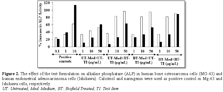
Assessment
of alkaline phosphatase (ALP) activity
The effect
of the test formulation on bone-specific alkaline phosphatase level is shown in
Figure 2. The positive control,
calcitriol showed 24.82%, 33.7% and 62.95% increase the level of ALP at 0.1, 1
and 10 nM, respectively in MG-63 cells. Moreover, the experimental groups
showed 90%, 87.3% and 86.9% increase the level of ALP in the UT-Med + BT-TI, BT-Med
+ UT-TI and BT-Med + BT-TI groups, respectively with respect to the UT-Med +
UT-TI group at 10 µg/mL. At 50 µg/mL, the percent ALP was significantly
increased by 81.4%, 79.6% and 70.6% in the UT-Med + BT-TI, BT-Med + UT-TI and
BT-Med + BT-TI groups, respectively compared to the UT-Med + UT-TI group (Figure 2). Besides, the positive
control naringenin showed 21.5%, 39.43% and 113.64% increase the level of ALP
at 0.1, 1 and 10 nM, respectively in Ishikawa cells. ALP percent was
significantly increased by 137% in the BT-Med + UT-TI group compared to the
UT-Med + UT-TI group at 1 µg/mL. Moreover, the experimental groups showed 32.8%
and 33.6% increase the level of ALP in the BT-Med + UT-TI and BT-Med + BT-TI
groups, respectively with respect to the UT-Med + UT-TI group at 10 µg/mL. At
50 µg/mL, the percent ALP was significantly increased by 46% in the BT-Med +
BT-TI group compared to the UT-Med + UT-TI group (Figure 2). The ALP plays a
vital role for the mineralization bone and considered a useful biochemical
marker for bone formation [44]. The bone biomarkers like bone alkaline
phosphatase (B-ALP) is considered to be a good marker for bone formation
released during the bone remodeling processes [45]. Combination with the
measurement of bone mineral density (BMD), the clinical applications of bone
biomarkers have provided comprehensive information for diagnosis of
osteoporosis [46]. In this experiment, the level of ALP was
revealed that the Biofield Energy Healing Treated test formulation significantly
increased the level of ALP expression, which might be very helpful to the
patients suffering from various bone-related disorders.
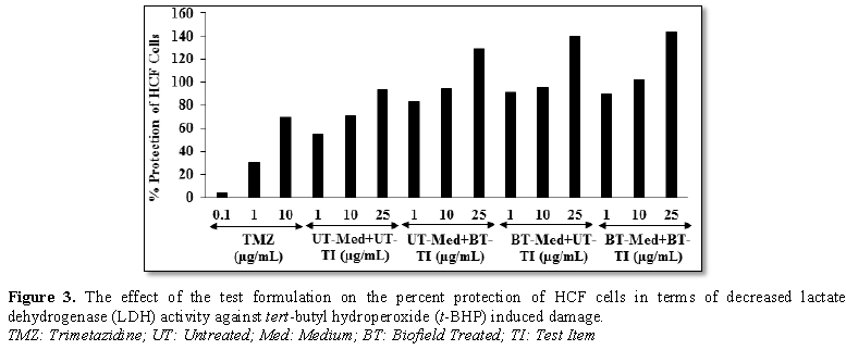
Estimation of
lactate dehydrogenase (LDH) activity in human cardiac fibroblasts (HCF)
The effect
of test items on the percent protection of HCF cells in terms of decreased
level of lactate dehydrogenase (LDH) activity is shown in Figure 3. The positive control, trimetazidine (TMZ) exhibited
3.59%, 30.14% and 69.42% protection of HCF cells (decreased of LDH activity)
compared to the t-BHP group. The
percent protection of HCF cells (decreased of LDH activity) was significantly
increased by 52.1%, 65.9% and 63.5% in the UT-Med + BT-TI, BT-Med + UT-TI and
BT-Med + BT-TI groups, respectively at 1 µg/mL as compared to the UT-Med +
UT-TI group. Moreover, at 10 µg/mL, the percent protection of HCF cells
(decreased of LDH activity) was significantly increased by 33.2%, 33.7% and
44.9% in the UT-Med + BT-TI, BT-Med + UT-TI and BT-Med + BT-TI groups,
respectively as compared to the UT-Med + UT-TI group. Further, percent
protection of HCF cells (decreased of LDH activity) was also significantly
increased by 37.7%, 49.8% and 53.3% in the UT-Med + BT-TI, BT-Med + UT-TI and
BT-Med + BT-TI groups, respectively at 25 µg/mL as compared to the UT-Med +
UT-TI group (Figure 3). The lactate
dehydrogenase (LDH) enzyme is mainly present in the heart and skeletal muscle.
It catalyzes the interconversion of pyruvate and lactate, which are critical
fuel metabolites of skeletal muscle particularly during exercise [47]. Elevated
serum lactic dehydrogenase (LDH) levels are associated with increased
cardiovascular mortality. Several inflammatory diseases were also correlated
with serum LDH [48]. The study results found that there was a significant
reduction of LDH level after Biofield Energy Treatment which protected heart
cells and might be helpful to resist against various pathological conditions
like tissue injury, necrosis, hemolysis or malignancies, hypoxia, etc. It also
indicates that the heart cells acted normally under stress and anaerobic
condition and improved overall heart function.
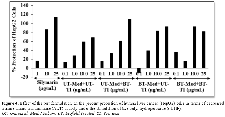
Estimation of alanine amino transferase (ALT) activity in HepG2 cells
The effect
of the test formulation on protection of HepG2 cells in terms of decrease
alanine amino transferase (ALT) activity is shown in Figure 4. The positive control, silymarin exhibited 16.35%, 85.83%
and 114.38% protection of HepG2 cells (decreased ALT activity). The protection
of HepG2 cells (decreased ALT activity) was significantly increased by 11% and
157% at 0.1 µg/mL in the UT-Med + BT-TI and BT-Med + BT-TI groups, respectively
as compared to the UT-Med + UT-TI group. Moreover, at 1 µg/mL, percent
protection of HepG2 cells (decreased ALT activity) was increased by 32.9% and
41.6% in the UT-Med + BT-TI and BT-Med + UT-TI groups, respectively as compared
to the UT-Med + UT-TI group. Additionally, protection of HepG2 cells (decreased
ALT activity) was also significantly increased by 41.8% and 58.9% in the BT-Med
+ UT-TI and BT-Med + BT-TI groups, respectively at 10 µg/mL as compared to the
UT-Med + UT-TI group. Further, the percent protection of HepG2 cells (decreased
ALT activity) was increased by 58.5%, 35.1% and 19.2% in the UT-Med + BT-TI,
BT-Med + UT-TI and BT-Med + BT-TI groups, respectively at 25 µg/mL as compared
to the UT-Med + UT-TI group (Figure 4).
Alanine Aminotransferase (ALT) activity plays as an indicator of general health
and especially on liver disorders. The probability of clinically significant
liver disease increases, particularly if the elevated ALT is associated with
symptoms such as fatigue, anorexia or pruritus [49]. In muscle, ALT plays an
important role for the regulation of glucose level during stressful conditions
such as fasting or vigorous exercise [50]. Here, the Biofield Energy Treatment
significantly protect liver hepatocytes in terms of reducing the level of
transaminases enzyme, ALT compared to the t-BHP
inducing group, which might be due to Consciousness Energy Healing Treatment to
the test formulation.
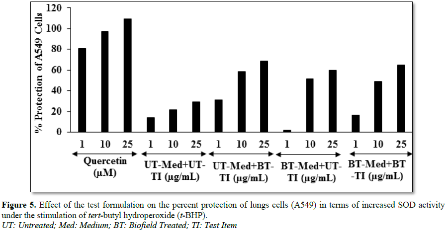
Estimation of
superoxide dismutase (SOD) activity in adenocarcinomic human alveolar basal
epithelial cells (A549)
The effect of the test formulation on the
protection of lungs cells (A549) in terms of increased super oxide dismutase
(SOD) activity is shown in Figure 5.
The positive control, showed 80.67%, 97.01% and 109.56% protection of A549
(lungs) cells (increased SOD activity) compared to the t-BHP group. The percent
protection of A549 (lungs) cells (increased SOD activity) was significantly
increased by 121.7% and 16.6% at 1 µg/mL in the UT-Med + BT-TI and BT-Med +
BT-TI groups, respectively compared to the UT-Med + UT-TI group. Moreover, at
10 µg/mL, the percent protection of A549 (lungs) cells (increased of SOD
activity) was significantly increased by 168.4%, 137% and 124% in the UT-Med +
BT-TI, BT-Med + UT-TI and BT-Med + BT-TI groups, respectively as compared to
the UT-Med + UT-TI group. Further, the level of SOD was increased by 135.4%,
103.7% and 121.1% in the UT-Med + BT-TI, BT-Med + UT-TI and BT-Med + BT-TI
groups, respectively at 25 µg/mL as compared to the UT-Med + UT-TI group (Figure 5). Extracellular SOD is one of
the three antioxidant enzyme isoforms, and is highly expressed in lungs and
vessels [51]. The lungs are directly exposed to more oxygen concentrations than
other tissues. Increase levels of both exogenous and endogenous reactive oxygen
species (ROS) leads to the pathogenesis of various lung disorders such as
asthma, chronic obstructive pulmonary disease (COPD), lung malignancies, etc.
[52]. Altogether, data observed that a significantly increased SOD levels after
Biofield Energy Treatment in A549 cells, were seen which might be helpful to
resist against various pathological conditions like oxidative stress and
related adverse effect. It also indicated that the lung cells acted normally
and improved overall respiratory activities.
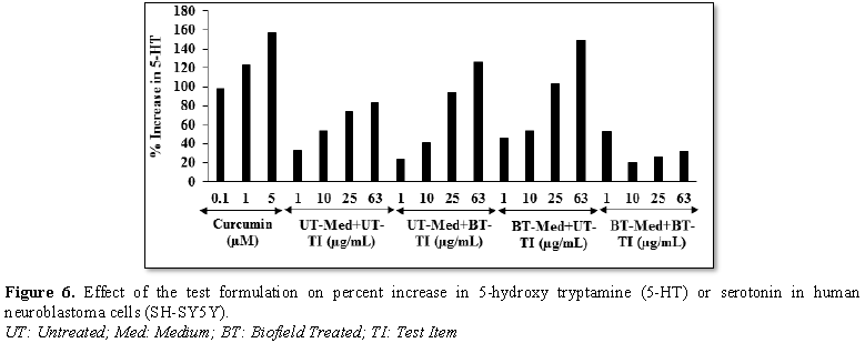
Effect
of test formulation on serotonin in human neuroblastoma (SH-SY5Y) cells
The effect of test formulation on serotonin level was assessed in SH-SY5Y
cells after 24 h of treatment by ELISA and the results are shown in Figure 6.
The positive control showed 98.2%, 123.53% and 156.76%
increase the level of serotonin. The level of serotonin was significantly
increased by 36.4% and 52.7% in the BT-Med + UT-TI and BT-Med + BT-TI groups,
respectively at 1 µg/mL compared to the UT-Med + UT-TI group. Moreover, at 10 µg/mL,
5-HT level was significantly increased by 19.7% in the BT-Med + BT-TI group as
compared to the UT-Med + UT-TI group. The serotonin level was significantly
increased by 27.5%, 40.2% and 26.6% in the UT-Med + BT-TI, BT-Med + UT-TI and
BT-Med + BT-TI groups, respectively at 25 µg/mL as compared to the UT-Med +
UT-TI group. Further, the serotonin level was significantly increased by 50.8%,
78.8% and 32.3% in the UT-Med + 52.7BT-TI, BT-Med + UT-TI and BT-Med + BT-TI
groups, respectively at 63 µg/mL as compared to the UT-Med + UT-TI group (Figure 6). Serotonin (5-HT) is a neurotransmitter
produced in neurons, gut, and heart cell mainly and responsible for stress,
anxiety, aggressive behavior and for the regulation of blood pressure [53]. Marazziti [54] demonstrated a clear view of
serotonergic dysfunction in different psychopathological disorders viz. schizophrenia, depression, anxiety
disorders, eating disorders, autism, and aggressive behaviors, etc. Therefore,
the data suggested that Biofield Energy Healing Treated novel test formulation
significantly improved the serotonin level, which would be highly useful
against various neurodegenerative diseases and other age-related disorders and
improved the normal functioning of the brain tissues.
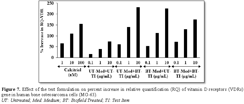
Effect of test
formulation on vitamin D receptors (VDRs)
Human bone osteosarcoma cells (MG-63) were treated with the test
formulation and the effect on VDR expression was determined using
quantitative-polymerase chain reaction (Q-PCR) amplification. VDR-relative
threshold cycle (VDR-CT) values were obtained from PCR amplification. Relative
quantification (RQ) was calculated from the VDR-CT and house-keeping (HK)-CT
values for MG-63 cells treated with test formulation and positive control is
represented in Figure 7. The positive control (calcitriol) showed
65.86%, 109.94% and 154.91% increase of RQ of VDR in a concentration-dependent
manner at 1, 10 and 100 nM, respectively. Moreover, RQ of VDR was significantly
increased by 265.5%, 219.5% and 335.3% in the UT-Med +
BT-TI, BT-Med + UT-TI and BT-Med
+ BT-TI groups, respectively at 0.1 µg/mL compared to the UT-Med + UT-TI group.
Additionally, at 1 µg/mL the VDR level was significantly increased by 253.4%,
185.9% and 228.7% in the UT-Med + BT-TI, BT-Med + UT-TI and BT-Med + BT-TI
groups, respectively compared to the UT-Med + UT-TI group. Further, VDR level
was also significantly increased by 212.7%, 203.5% and 136.3% in the UT-Med +
BT-TI, BT-Med + UT-TI and BT-Med + BT-TI groups, respectively at 10 µg/mL
compared to the UT-Med + UT-TI group. The role of vitamin D with extra-skeletal
system like autoimmune disease, cardiovascular disease and cancer is of major
interest nowadays [58]. Absence of a functional VDR or the key activating
enzyme, 25-OHD-1α-hydroxylase (CYP27B1), causes congenital disease or severe
vitamin D deficiency. Deficiency of vitamin D in humans is associated with
increased prevalence of diseases [55]. Overall, the Consciousness Energy
Healing Treated test formulation has tremendously increased the expression of
VDRs, which might be helpful to bind more active vitamin D3
metabolites and that ultimately can improve the more physiological functions of
vitamin D and simultaneously improved bone cell growth and development.
CONCLUSION
The study results
showed that the novel test formulation was safe and non-toxic based on MTT cell
viability assay in six tested cells. The BT-Med + BT-TI group showed 181% and
82.2% restoration of cell viability at 1 and 10 µg/mL, respectively in human cardiac
fibroblasts cells (HCF) compared to the UT-Med + UT-TI group. Moreover, the
UT-Med + BT-TI group showed 126.8% and 86.3% restoration of cell viability at
10 and 25 µg/mL, respectively in human hepatoma cells (HepG2) compared to the
untreated group. Additionally, 101.2% (at 10 µg/mL), 103.6% (at 10 µg/mL) and
135% (at 25 µg/mL) restoration of cell viability was observed in
adenocarcinomic human alveolar basal epithelial cells (A549) by UT-Med + BT-TI,
BT-Med + UT-TI and BT-Med + BT-TI groups, respectively compared to the
untreated group. Alkaline phosphatase (ALP) activity was significantly
increased by 90% and 87.3% in the UT-Med + BT TI and BT-Med + UT TI groups,
respectively at 10 µg/mL in human bone osteosarcoma cells (MG-63). Moreover,
ALP activity was significantly increased by 137% in the BT-Med + UT-TI group at
1 µg/mL than untreated group. The percent protection of HCF cells (decreased
LDH activity) was significantly increased by 65.9% and 63.5% at 1 µg/mL in the
BT-Med + UT-TI and BT-Med + BT-TI groups, respectively compared to the
untreated. The percent protection of HepG2 cells (decreased ALT activity) was
significantly increased by 157% at 0.1 µg/mL in the BT-Med + BT-TI group
compared to the untreated group. The percent protection of A549 (lungs) cells
(increased SOD activity) was significantly increased by 168% and 135.4% in the
UT-Med + BT-TI group at 10 and 25 µg/mL, respectively compared to the untreated
group. The serotonin level was significantly increased by 78.8% (at 63 µg/mL)
and 52.7% (at 1 µg/mL) in the BT-Med + UT-TI and BT-Med + BT-TI groups,
respectively compared to the untreated group in human neuroblastoma cells
(SH-SY5Y). The relative quantification (RQ) of vitamin D receptors (VDRs) level
was significantly increased by 265.5% (at 0.1 µg/mL) and 253.4% (at 1 µg/mL) in
the UT-Med + BT-TI group; however, 335.3% in the BT-Med + BT-TI group at 0.1
µg/mL compared to the untreated group in MG-63 cells. In conclusion, the
Biofield Energy Treatment significantly improved heart, liver, bones, neuronal
and lungs related biomarkers and also protected cardiomyocyte, hepatocyte,
osteocytes, pneumocyte and nerve cells from oxidative damage induced by tert-butyl hydroperoxide (t-BHP). Thus, the results suggest that
Biofield Energy Treatment can be used as a complementary and alternative
treatment for the prevention of various types of cardiac disorders (peripheral
artery disease, high blood pressure, congenital heart disease, stroke,
congestive heart failure, rheumatic heart disease, carditis, valvular heart disease,
thromboembolic disease and venous thrombosis, etc.), hepatic disorders
(cirrhosis, Wilson disease, liver cancer, hemochromatosis) and lungs disorders
(asthma, emphysema, chronic bronchitis, pneumonia, cystic fibrosis). Further,
it can be useful to improve cell-to-cell messaging, normal cell growth and
differentiation, cell cycling and proliferation, neurotransmission, skin
health, hormonal balance, immune and cardiovascular functions. Moreover, it can
also be utilized in organ transplants (i.e., liver, kidney and heart
transplants), aging, hormonal imbalance and various inflammatory and
immune-related disease conditions like Alzheimer’s Disease (AD), Dermatitis,
Asthma, Ulcerative Colitis (UC), Hashimoto Thyroiditis, Pernicious Anemia,
Sjogren Syndrome, Aplastic Anemia, Multiple Sclerosis, Hepatitis, Graves’
Disease, Irritable Bowel Syndrome (IBS), Dermatomyositis, Diabetes, Myasthenia
Gravis, Atherosclerosis, Parkinson’s Disease, Systemic, etc., to Lupus
Erythematosus (SLE), stress, improve overall health and Quality of Life.
ACKNOWLEDGEMENT
Authors gratefully acknowledged to Trivedi
Global, Inc., Trivedi Science and Trivedi Master Wellness for their support. In
addition, authors are thankful for the support of Dabur Research Foundation for
conducting this study.
1.
Global Burden of Disease Collaborative Network
(2017) Global Burden of Disease Study 2016 (GBD 2016) Results. Seattle, United
States: Institute for Health Metrics and Evaluation (IHME).
2.
Pal S (2018) Incidence and prevalence of major
neurologic disorders. US Pharm 43: 24.
3.
Rakesh S, Arunporn I (2017) Herbal supplements or
herbs in heart disease: Herbiceutical formulation, clinical trials, futuristic
developments. J Cardiol Cardiovasc Ther 3: 555603.
4.
Contreras-Zentella ML, Hernández-Muñoz R (2016) Is
liver enzyme release really associated with cell necrosis induced by oxidant
stress? Oxid Med Cell Longev 2016: 3529149.
5.
Schmidt E, Schmidt FW (1970) Aspects of enzyme
diagnosis. Med Welt 21: 805-816.
6.
Frederiks WM, Vogels IM, Fronik GM (1984) Plasma
ornithine carbamyl transferase level as an indicator of ischemic injury of rat
liver. Cell Biochem Funct 2: 217-220.
7.
Boots AW, Haenen GR, Bast A (2003) Oxidant
metabolism in chronic obstructive pulmonary disease. Eur Respir J 46: 14-27.
8.
Romieu I (2005) Nutrition and lung health. Int J
Tuberc Lung Dis 9: 362-374.
9.
Kelly FJ (2005) Vitamins and respiratory disease:
Antioxidant micronutrients in pulmonary health and disease. Proc Nutr Soc 64:
510-526.
10.
Fischer AG, Ullsperger M (2017) An update on the
role of serotonin and its interplay with dopamine for reward. Front Hum
Neurosci 11: 484.
11.
Anonymous (2006) A sensible 10-year plan for mental
health. Lancet 367: 86.
12.
Ryan-Harshman M, Aldoori W (2005) Health benefits
of selected minerals. Can Fam Physician 51: 673-675.
13.
Rayman MP (2000) The importance of selenium to
human health. Lancet 356: 233-241.
14.
Beard JL, Connor JR (2003) Iron status and neural
functioning. Ann Rev Nutr 23: 41-58.
15.
Coleman CI, Hebert JH, Reddy P (2003) The effects
of Panax ginseng on quality of life. J Clin Pharm Ther 28: 5-15.
16.
Das L, Bhaumik E, Raychaudhuri U, Chakraborty R
(2011) Role of nutraceuticals in human health. J Food Sci Technol 49:173-183.
17.
Lutgendorf SK, Mullen-Houser E, Russell D, Degeest
K, Jacobson G et al. (2010) Preservation of immune function in cervical cancer
patients during chemoradiation using a novel integrative approach. Brain Behav
Immun 24: 1231-1240.
18.
Ironson G, Field T, Scafidi F, Hashimoto M, Kumar M
et al. (1996) Massage therapy is associated with enhancement of the immune
system's cytotoxic capacity. Int J Neurosci 84: 205-217.
19.
Jain S, Hammerschlag R, Mills P, Cohen L, Krieger R
et al. (2015) Clinical studies of biofield therapies: Summary, methodological
challenges, and recommendations. Glob Adv Health Med 4: 58-66.
20.
Rubik B (2002) The biofield hypothesis: Its
biophysical basis and role in medicine. J Altern Complement Med 8: 703-717.
21.
Trivedi MK, Patil S, Shettigar H, Mondal SC, Jana S
(2015) The potential impact of biofield treatment on human brain tumor cells: A
time-lapse video microscopy. J Integr Oncol 4: 141.
22.
Trivedi MK, Patil S, Shettigar H, Gangwar M, Jana S
(2015) In vitro evaluation of biofield treatment on cancer biomarkers
involved in endometrial and prostate cancer cell lines. J Cancer Sci Ther 7:
253-257.
23.
Trivedi MK, Tallapragada RM (2008) A transcendental
to changing metal powder characteristics. Met Powder Rep 63: 22-28, 31.
24.
Trivedi MK, Nayak G, Patil S, Tallapragada RM,
Latiyal O (2015) Studies of the atomic and crystalline characteristics of
ceramic oxide nanopowders after bio field treatment. Ind Eng Manage 4: 161.
25.
Trivedi MK, Branton A, Trivedi D, Nayak G, Charan S
et al. (2015) Phenotyping and 16S rDNA analysis after biofield treatment on Citrobacter
braakii: A urinary pathogen. J Clin Med Genom 3: 129.
26.
Trivedi MK, Patil S, Shettigar H, Mondal SC, Jana S
(2015) An impact of biofield treatment: Antimycobacterial susceptibility
potential using BACTEC 460/MGIT-TB System. Mycobact Dis 5: 189.
27.
Trivedi MK, Branton A, Trivedi D, Nayak G, Mondal
SC et al. (2015) Morphological characterization, quality, yield and DNA
fingerprinting of biofield energy treated alphonso mango (Mangifera indica
L.). Journal of Food and Nutrition Sciences 3: 245-250.
28.
Trivedi MK, Branton A, Trivedi D, Nayak G, Mondal
SC et al. (2015) Evaluation of biochemical marker – Glutathione and DNA
fingerprinting of biofield energy treated Oryza sativa. Am J Biosci 3: 243-248.
29.
Trivedi MK, Branton A, Trivedi D, Nayak G, Plikerd
WD et al. (2017) A Systematic study of the biofield energy healing treatment on
physicochemical, thermal, structural and behavioral properties of magnesium
gluconate. Int J Bioorg Chem 2: 135-145.
30.
Parulkar VR, Trivedi MK, Branton A, Trivedi D,
Nayak G et al. (2018) Improved metabolism of vitamin D3 in human osteoblasts
cells after biofield energy healing treatment. Am J Lab Med 3: 11-19.
31.
Trivedi MK, Patil S, Shettigar H, Bairwa K, Jana S
(2015) Phenotypic and biotypic characterization of Klebsiella oxytoca:
An impact of biofield treatment. J Microb Biochem Technol 7: 203-206.
32.
Nayak G, Altekar N (2015) Effect of biofield
treatment on plant growth and adaptation. J Environ Health Sci 1: 1-9.
33.
Branton A, Jana S (2017) The influence of energy of
consciousness healing treatment on low bioavailable resveratrol in male Sprague
Dawley rats. Int J Clin Dev Anatomy 3: 9-15.
34.
Branton A, Jana S (2017) The use of novel and
unique biofield energy healing treatment for the improvement of poorly
bioavailable compound, berberine in male Sprague Dawley rats. Am J Clin Exp Med
5: 138-144.
35.
Branton A, Jana S (2017) Effect of The biofield
energy healing treatment on the pharmacokinetics of 25-hydroxyvitamin D3
[25(OH)D3] in rats after a single oral dose of vitamin D3. Am J Pharmacol
Phytother 2: 11-18.
36.
Parulkar VR, Trivedi MK, Branton A, Trivedi D,
Nayak G et al. (2017) The use of consciousness energy healing based
herbomineral formulation for skin anti-aging strategies. J Food Nutr Sci 5:
96-106.
37.
Singh J, Trivedi MK, Branton A, Trivedi D, Nayak G
et al. (2017) Consciousness energy healing treatment based herbomineral
formulation: A safe and effective approach for skin health. Am J Pharmacol
Phytother 2: 1-10.
38.
Anagnos D, Trivedi K, Branton A, Trivedi D, Nayak G
et al. (2018) Influence of biofield treated vitamin D3 on proliferation,
differentiation, and maturation of bone-related parameters in MG-63 cell-line.
Int J Biomed Eng Clin Sci 4: 6-14.
39.
Lee AC, Trivedi K, Branton A, Trivedi D, Nayak G et
al. (2018) The potential benefits of biofield energy treated vitamin D3 on bone
mineralization in human bone osteosarcoma cells (MG-63). Int J Nutr Food Sci 7:
30-38.
40.
Stutheit ME, Trivedi K, Branton A, Trivedi D, Nayak
G et al. (2018) Biofield energy treated vitamin D3: Therapeutic implication on
bone health using osteoblasts cells. Am J Life Sci 6: 13-21.
41.
Rubio V, García-Pérez AI, Tejedor MC, Herráez A,
Diez CJ (2017) Esculetin neutralises cytotoxicity of t-BHP but not of H2O2
on human leukemia NB4 cells. BioMed Res Int Article ID 9491045: 9.
42.
Nemzer B, Chang T, Xie Z, Pietrzkowski Z, Reyes T,
et al. (2014) Decrease of free radical concentrations in humans following
consumption of a high antioxidant capacity natural product. Food Sci Nutr 2:
647-654.
43.
Örs G, Gülçe Iz S (2018) Cytoprotective effect of a
functional anti-pollutant blend through reducing B[a] P-induced intracellular
oxidative stress and UVA exposure. Turk J Biol 42: 453-462.
44.
Orimo HJ (2010) The mechanism of mineralization and
the role of alkaline phosphatase in health and disease. Nippon Med Sch 77:
4-12.
45.
Tobiume H, Kanzaki S, Hida S, Ono T, Moriwake T, et
al.(1997) Serum bone alkaline phosphatase isoenzyme levels in normal children
and children with growth hormone (GH) deficiency: A potential marker for bone
formation and response to GH therapy. J Clin Endocrinol Metab 82: 2056-2061.
46.
Mukaiyama K, Kamimura M, Uchiyama S, Ikegami S,
Nakamura Y, et al. (2015) Elevation of serum alkaline phosphatase (ALP) level
in postmenopausal women is caused by high bone turnover. Aging Clin Exp Res 27:
413-418.
47.
Liang X, Liu L, Fu T, Zhou Q, Zhou D, et al. (2016)
Exercise inducible lactate dehydrogenase B regulates mitochondrial function in
skeletal muscle. J Biol Chem 291: 25306-25318.
48.
Wu LW, Kao TW, Lin CM, Yang HF, Sun YS (2016)
Examining the association between serum lactic dehydrogenase and all-cause
mortality in patients with metabolic syndrome: A retrospective observational
study. BMJ Open 6: e011186.
49.
Kim WR, Flamm SL, Di Bisceglie AM, Bodenheimer HC
(2008) Serum activity of alanine aminotransferase (ALT) as an indicator of
health and disease. Hepatology 47: 1363-1370.
50.
Mc Commis KS, Chen Z, Fu X, Mc Donald WG, Colca JR,
et al. (2015) Loss of mitochondrial pyruvate carrier 2 in the liver leads to
defects in gluconeogenesis and compensation via pyruvate-alanine cycling. Cell
Metab 22: 682-694.
51.
Kinnula VL, Crapo JD (2003) Superoxide dismutases
in the lung and human lung diseases. Am J Respir Crit Care Med 167: 1600-1619.
52.
Kliment CR, Tobolewski JM, Manni ML, Tan RJ,
Enghild J, et al. (2008) Extracellular superoxide dismutase protects against
matrix degradation of heparan sulfate in the lung. Antioxid Redox Signal 10:
261-8.
53.
Dorszewska J, Prendecki M, Oczkowska A, Rozycka A,
Lianeri M, et al. (2013) Polymorphism of the COMT, MAO, DAT, NET and 5-HTT
genes and biogenic amines in Parkinson’s disease. Curr Genomics 14: 518-533.
54.
Marazziti D (2017) Understanding the role of
serotonin in psychiatric diseases. F1000Res 6: 180.
55.
Yamamoto Y, Yoshizawa T, Fukuda T, Shirode-Fukuda
Y, Yu T, et al. (2013) Vitamin D receptor in osteoblasts is a negative
regulator of bone mass control. Endocrinology 154: 1008-1020.
-
 Table 1
QUICK LINKS
- SUBMIT MANUSCRIPT
- RECOMMEND THE JOURNAL
-
SUBSCRIBE FOR ALERTS
RELATED JOURNALS
- Advance Research on Alzheimers and Parkinsons Disease
- Chemotherapy Research Journal (ISSN:2642-0236)
- Journal of Pathology and Toxicology Research
- Archive of Obstetrics Gynecology and Reproductive Medicine (ISSN:2640-2297)
- Journal of Cancer Science and Treatment (ISSN:2641-7472)
- International Journal of Radiography Imaging & Radiation Therapy (ISSN:2642-0392)
- Journal of Infectious Diseases and Research (ISSN: 2688-6537)


