1419
Views & Citations419
Likes & Shares
Background: Uterine
Leiomyoma is a benign neoplasm that occurs from the overgrowth of smooth muscle
and connective tissue in the uterus. A nuclear medicine scan is a sensitive
indicator of metastatic bone diseases or other pathology. The diagnosis of
uterine leiomyomas using nuclear medicine is straightforward, given the common
clinical manifestations and typical imaging features.
Case presentation: A 45-year-old
woman with a history of nasopharyngeal cancer was referred for a bone scan to
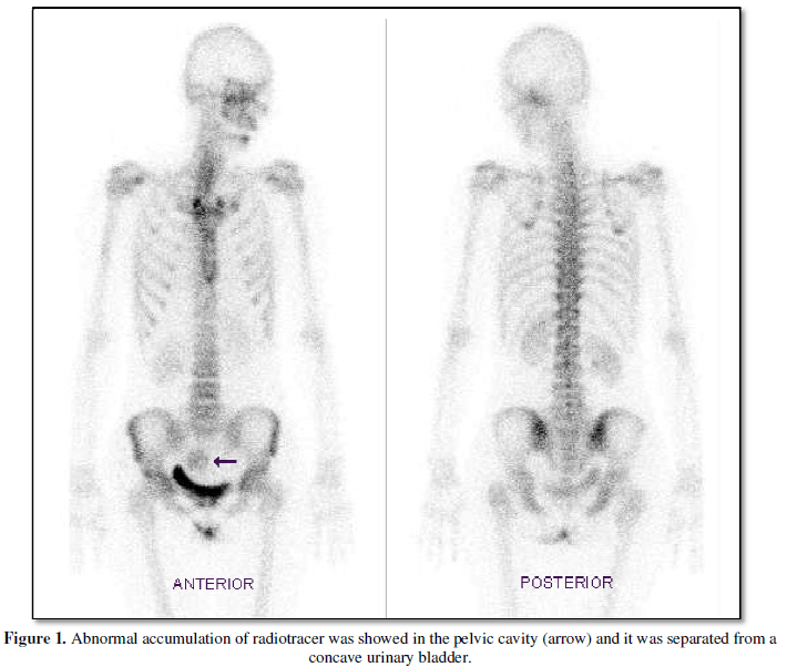
assess for bone metastasis. Abnormal accumulation of radiotracer was shown in
the pelvic cavity.
Conclusion: The differential
diagnosis of the abnormal radioactivity included a sacral metastasis and a
pelvic tumor with uptake of bone-seeking tracers. The ultrasonography of the
pelvic cavity showed a huge uterine myoma with calcifications and cystic
components. After the testing, a total abdominal hysterectomy was performed and
multiple leiomyomata were confirmed by histopathologic examination.
Keywords: Bone Scan, Uterine Leiomyoma, Pelvic Tumor
INTRODUCTION
Uterine leiomyoma has been reported as an occasional cause of
extraskeletal uptake of a bone-seeking agent in the pelvis [1-5]. The cause of
tumor uptake of bone-seeking tracers is thought to be related to dystrophic
calcification [6]. In dystrophic calcification, the mechanism appears to be the
loss of intracellular calcium in the injured cells resulting in an increased
calcium-binding capacity.
CASE REPORT
The bone scan was performed three hours after an intravenous injection of 740MBq (20mCi) Tc-99m methylene diphosphonate (MDP) into a 45-year-old woman with a history of nasopharyngeal cancer. Abnormal accumulation of radiotracer was shown in the pelvic cavity (arrow) and it was separated from a concave urinary bladder (Figure 1). The differential diagnosis of the abnormal radioactivity included sacral metastasis and a pelvic tumor with uptake of bone-seeking tracers. The patient presented with vaginal spotting, dyspareunia, dysmenorrhea, and urinary urgency. Physical examination showed pale conjunctiva and a palpable mass in the lower abdomen. Laboratory data revealed a reduction of hemoglobin to 8.6 g/dl (normal range, 12 to 14 g/dl).
Then ultrasonography was arranged to evaluate the pelvic mass, which
demonstrated a heterogeneous hypo-echoic mass with calcifications and cystic
components. It originated from the uterus and was approximately 11.0 x 10.0 x
9.7 cm in size (Figure 2). The
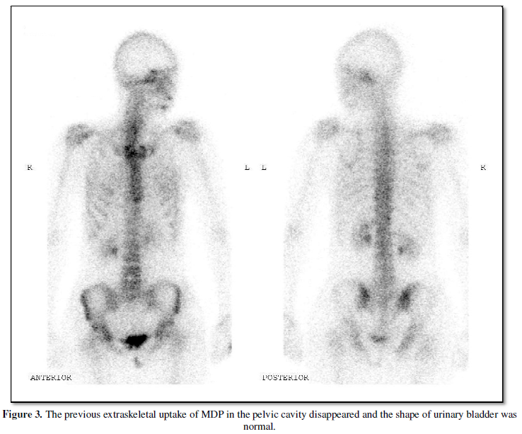
patient underwent total abdominal hysterectomy for the uterine mass. The uterus
measured 16.6 x 12.1 x 7.1 cm and weighed 801 gm. The histopathologic diagnosis
revealed multiple intramural and subserous leiomyomata of the uterus. The
largest measured up to 7 cm in diameter. The bone scan was repeated after the
hysterectomy. The previous extraskeletal uptake of MDP in the pelvic cavity
disappeared and the shape of the urinary bladder was normal (Figure 3).
DISCUSSION
Incidental extraskeletal uptake of bone-seeking tracers on bone scans
is occasionally seen. Soft tissue uptake must be differentiated from bone
pathology [7]. Additionally, normal renal uptake and excretion of bone-seeking
agents allow for gross visualization of the genitourinary system and provide
useful diagnostic information. Retroperitoneal or pelvic masses should be
suspected if abnormalities in the location or contour of urinary bladder are
present [7-11]. Obtaining a relevant clinical history and laboratory data is
helpful to clarify the incidental extraskeletal uptake of bone-seeking tracers.
COMPETING
INTERESTS
The authors declare they have no conflict of interests in publishing
this case study.
ACKNOWLEDGMENTS
This work was supported by
Research Grants MOST 103-2320-B-037-025 from the Ministry of Science and
Technology, KMU-TP105E12, KMU-TP105PR06, KMU-M106029, 105KMUOR02 and
KMU-O104003 (Aim for the Top 500 Universities Grant) from Kaohsiung Medical
University, and NSYSUKMU106-P011 from NSYSU-KMU Research Project, Taiwan.
1. Ell PJ, Breitfellner G, Meixner M.
Technetium-99m-HEDP concentration in calcified myoma. J Nucl Med. 1976;
17:323-324.
2. Stadalnik RC. Non-skeletal accumulation of
bone seeking agents: pelvis. Sem Nucl Med. 1988; 18:159-161.
3. Rohrer DG,
Williamson BR, Teates CD. Calcified uterine
leiomyoma simulating metastatic disease on bone scan. South Med J. 1988;
81:651-652.
4. Inoue H, Aizawa N, Mizuno T, et al. A large
degenerated subserous leiomyoma of the uterus: uncommon scintigraphic and
ultrasonographic findings. Ann Nucl Med. 1989; 3:55-57.
5. Buyukdereli G, Guney IB, Kibar M. Bone scan
findings of sacrococcygeal chordoma and uterine leiomyoma. J Women’s Imaging.
2004; 6:171-174.
6. Elgazzar A. H. Orthopedic Nuclear Medicine.
Berlin Heidelberg. Springer-Verlag, 2004, pp197-198.
7. Loutfi I, Collier BD, Mohammed AM.
Nonosseous abnormalities on bone scans. J Nucl Med Technol. 2003; 31:149-153.
8. Shanley DJ,
9. Ackerman L, Shirazi P, Van Drunen M.
Bladder displaced by stool-distended rectosigmoid colon presenting on bone scan
as a pelvic soft tissue mass. Clin Nucl Med. 1997; 22:733-734.
10. Al Naeem AN, Powe J, Bakheet S, et al.
Displaced urinary bladder creating an unusual pattern on bone scan mimicking
disease. Clin Nucl Med. 1999; 24:137-138.
11. Lin ST, Sohn MH, Park SA. Curious
radioactivity in the lower abdomen on bone scintigraphy: displacement of the
urinary bladder by an incidentally diagnosed uterine myoma. Clin Nucl Med.
2000; 25:824-825.

QUICK LINKS
- SUBMIT MANUSCRIPT
- RECOMMEND THE JOURNAL
-
SUBSCRIBE FOR ALERTS
RELATED JOURNALS
- BioMed Research Journal (ISSN:2578-8892)
- International Journal of Internal Medicine and Geriatrics (ISSN: 2689-7687)
- Journal of Ageing and Restorative Medicine (ISSN:2637-7403)
- Journal of Rheumatology Research (ISSN:2641-6999)
- Journal of Nursing and Occupational Health (ISSN: 2640-0845)
- Journal of Allergy Research (ISSN:2642-326X)
- Journal of Blood Transfusions and Diseases (ISSN:2641-4023)
")
")


