803
Views & Citations10
Likes & Shares
Objective: Designing virtual centers and pathways of the brain and spine for education.
Methodology: With NVidia software we designed immersive stereoscopic software to navigate in 3D through a virtual brain and spine cords.
Results: 33 centers, 7 pathways and 20 tracts of the central nervous system (CNS) have been developed on a basic model of brain anatomy. A Cyber Brain 3D VR System was designed.
Conclusion: Education is the main objective of this program which will aloud a general understanding of the anatomy and physiology of the CNS. We are proposing for it to be used by medical students as a virtual tool and with patients for a better understanding of clinical explanations. This immersive virtual scenario is available for 3D glasses and potentially for Oculus rift + Touch.
Keywords: Brain, Virtual reality, Virtual simulator
INTRODUCTION
Surgical education
There are two different ways of seeing anatomy; one in the real and one in the virtual world. The first one includes cadaveric and animals dissection, open and endoscopic surgery, endoscopy, and robotic surgery (immersive anatomy with Da Vinci robot). Virtual anatomy includes books, e.g. Ullan, Kierman and Lopez’s books [1-3], journals, image in 2D, image in 3D and augmented reality. The way people perceive the anatomy varies depending on the media in which it is displayed. All methods are necessary and at the same time complementary. The ways people learn vary from individual to individual, some learners prefer visual stimuli and feedback controllers as in an open surgery or augmented reality with total immersion in which natural or artificial feedback systems use many senses for learning.
Patient’s education
More and more often patients are aware of their conditions and look for different sources to learn more about the, they get information from the internet and they review images, videos and texts about their conditions. With this technology we can facilitate to our patients the understanding of medical condition by showing them virtual scenarios. The information can include the etiology, symptoms, treatments and prognosis of their diseases and would replace the use of posters, magazines, drawings and other 2D media.
There are a lot of brain and spine cord virtual scenarios [4-6], but the particular difference between them and ours is the number of nuclei, and tracts displayed. These characteristics help to understand in. an easier way the neuroanatomy and cranial centers in the brainstem and 13 nuclei of the spine cord and ascending and descending tracts. Our design the neurophysiology, for instance, we included an amygdaline body with 6 nuclei, all hypothalamus nuclei, 12 permits the user to navigate through specific neurological pathways, look inside of the hemispheres and contemplate how the mind works. We are using this software as a complementary tool in the psychological treatment of obese patients [7-9].
Obesity pathophysiology includes structures such as the hypothalamus, limbic system and prefrontal cortex that affect the hunger and satiety functions.
MATERIALS AND METHODS
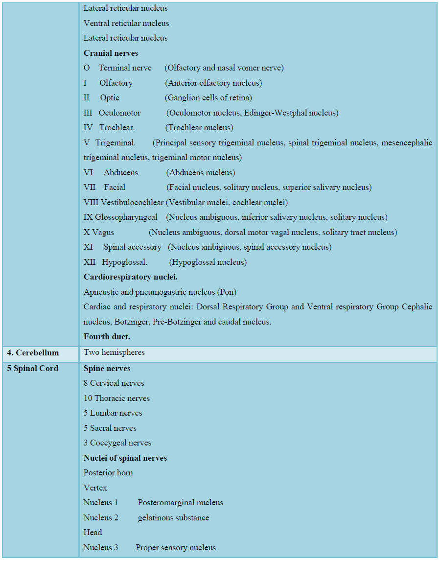
The basic model of the brain includes cerebral cortex with hemispheres (frontal, temporal, parietal and occipital lobes), diencephalon, midbrain, pons, cerebellum, brainstem, eyeballs, olfactory, visual nerves and ventricular system (lateral and III-IV ventricles as well as the mesencephalic duct). The NVidia GeForce GT 430 card was used as the platform. The program used to design the structures of the brain was 3D game studio; the virtual models were made on 3D Max. The following structures (Table 1) were obtained from 4 Neuroanatomical books.
We used a PC processor with Intel Core i5-760 2.8 GHz, Windows 7 Home Premium de 64 bits. Memory SDRAM DDR3 4 GB, a hard disc 500 GB SATA II 7200 RPM. Logitech LS11 stereo sound 2.0 and a sound card Genius SM-Value 5.1 channel. User can display the images on a screen, wall or use Head Mounted Display eMagin with a Benq 3D active shutter glasses for DLP link ready projector (Projector benq MX710/MX711/MX762ST series, supported timing for HMDI [HDCP] input with 1024 × 768 XGA resolution and 3200 lumens).
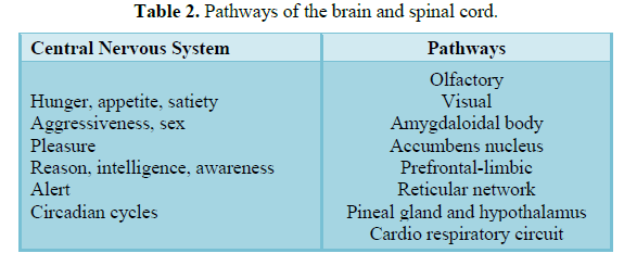
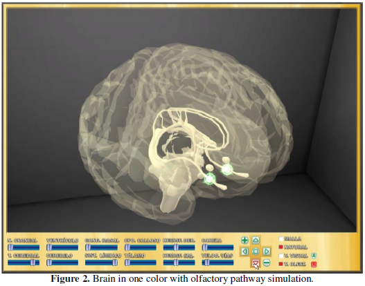
There is a virtual keyboard (Figure 1) to move the brain on the screen, there are 5 arrows to move up, down, right and left the screen, and another one to zoom in. Six bars to make solid or transparent the following structures: right hemisphere, left hemisphere, corpus callosum, ventricles, diencephalon, midbrain, pons, cerebellum, brainstem, thalamus, cranial nerves, and basal ganglia. Another bars located to the right of the screen permits users to run pathways including the olfactory, visual, limbic, amygdaloid body, accumbens nucleus, prefrontal and limbic, reticular network, pineal, hypothalamus and lateral spinothalamic tract. On any type of display the brain can be seen in colors or only in white, is possible to dissect and disappear superficial structures to recognize deeper ones (Figure 2 and Table 2).
RESULTS
A Cyber brain 3D VR System™ with a turnkey system with stereoscopic 3D visualization software was developed. Software and hardware were created, including a 3D computer, 3D projector, and the 3D glasses. Table 1 describes the CNS structures represented in the software.
DISCUSSION AND CONCLUSION
Cyber Brain 3D VR System™ includes a turnkey system with stereoscopic 3D visualization software that represents an alternative tool for medical education. However, we can suggest other applications using the system as patient’s education. Neuroanatomy is so brad that goes beyond the spectrum of this project but this atlas includes the most important structures in the central nervous system. Is important to notice that anatomy perspective varies among the different authors. We propose to use this system in high school and undergraduate students as a complementary tool to neuroanatomy books, journals, webpages, and videos. In medical school, cadaver dissection facilitates learning in neuroanatomy but remains a tough subject. Our software will help solidifying students’ knowledge after dissection. Actually as of today, medical students from Universidad Panamericana, School of Medicine, the top medical school in Mexico, are using this system as a complementary toolkit after cadaveric dissections. As mentioned before, patients can use this system to get a better understanding of their neurological diseases, and can aloud neurosurgeons to teach patients about their surgical procedures. This is a useful tool for psychiatrists and psychologists as well; they can explain to their patients how the mind works, what is the relationship between structures of the brain and their behavior, emotions, intelligence, memory and consciousness. Addictions and obesity are two of the most common diseases that can be explained using out system. For all the above, stereoscopic immersion, animation and simulation make learning easier, funnier and more interesting. A journey through the human brain allows all our senses to be involved in the understanding and learning process [10-18]. In future endeavors, this system could be improved with feedback tools like cyber gloves or Kinect using big screens as virtual cubes for total immersion; oculus rift + touch is the best option up today [19-21].
1. Ullan-Serrano J (2006) Neuro-anatomy. 3rd Edn.
2. Barr ML, Kierman JA (1986) The human nervous system. Anatomic point of view. 4th Edn. Editorial Harala.
3. Lopez Antunez L (2003) Functional anatomy of the nervous system. Limusa, Noriega editors, Mexico.
4. Brewer DN, Wilson TD, Eagleson R, De Ribaupierre S (2012) Evaluation of neuroanatomical training using a 3D visual reality model. Stud Health Technol Inform 173: 85-91.
5. Debarba HG, Grandi J, Maciel A (2012) Anatomic hepatectomy planning through mobile display visualization and interaction. Stud Health Technol Inform 173: 111-115.
6. Sourin A, Yasmin S (2012) Haptic editing of MRI brain data. Stud Health Technol Inform 173: 490-496.
7. Ministry of Health (Secretaria de Salud, INSP) (2006) Prevalence of overweight and obesity in Mexico. Encuesta Nacional de Salud y Nutricion. Proceso INSP.
8. Coutiño JC (2007) Integral obesity treatment. Official Mexican Standard, pp: 578-587.
9. Hedman L, Fahlstedt M, Schlikum M (2012) Training diagnosis and treatment of cervical spine trauma using a new educational program for visualization through imaging and simulation (VIS): A first evaluation by medical students. In: Stud. Health Technol. Informatics, pp: 171-174.
10. Conte G, Ye A, Forbes A, Leow A (2015) BRAINtrinsic: A virtual reality-compatible tool for exploring intrinsic topologies of the human brain connectome. Lecture Notes in Computer Science 9250: 67-76.
11. Xia M, Wang J, He Y (2013) Brainnet viewer: A network visualization tool for human brain connectomics. PLoS One 8: e68910.
12. Sporns O, Tononi G, Kotter R (2005) The human connectome: A structural description of the human brain. PLoS Comput Biol 1: e42.
13. Sporns O (2011) The human connectome: A complex network. Ann N Y Acad Sci 1224: 109-125.
14. La Plante RA, Douw L, Tang W, Stufflebeam SM (2014) The connectome visualization utility: Software for visualization of human brain networks. PLoS One 9: e113838.
15. Forbes A, Villegas J, Almryde KR, Plante E (2014) A stereoscopic system for viewing the temporal evolution of brain activity clusters in response to linguistic stimuli. Proc SPIE Int Soc Opt Eng 9011: 90110I.
16. de Ridder M, Klein K, Kim J (2015) CereVA - Visual analysis of function brain connectivity. IVAPP 2015 - 6th International Conference on Information Visualization Theory and Applications; VISIGRAPP, Proceedings, pp: 131-138.
17. National Institute of Health (2015) The Human Connectome Project. Available at: http://www.humanconnectomeproject.org/
18. Human Brain Project (2015) Human Brain Project. Available at: https://www.humanbrainproject.eu/
19. Desai PR, Desai PN, Ajmera KD, Mehta K (2014) Review paper on oculus rift - A virtual reality headset. Int J Eng Trends Technol 13: 175-179.
20. Davis S, Nesbitt K, Nalivaiko E (2015) Comparing the onset of cyber sickness using the oculus rift and two virtual roller coasters. In: Proceedings of the 11th Australasian Conference on Interactive Entertainment (IE 2015), Sydney, Australia 167: 3-14.
-
 Table 1
 -
Table 2
-
Table 3
-
Table 4


QUICK LINKS
- SUBMIT MANUSCRIPT
- RECOMMEND THE JOURNAL
-
SUBSCRIBE FOR ALERTS
RELATED JOURNALS
- Oncology Clinics and Research (ISSN: 2643-055X)
- Journal of Spine Diseases
- Stem Cell Research and Therapeutics (ISSN:2474-4646)
- Dermatology Clinics and Research (ISSN:2380-5609)
- International Journal of Clinical Case Studies and Reports (ISSN:2641-5771)
- Journal of Cardiology and Diagnostics Research (ISSN:2639-4634)
- Journal of Clinical Trials and Research (ISSN:2637-7373)


