2065
Views & Citations1065
Likes & Shares
Epidermal nevi are hamartomatous proliferations of the epithelium.
We report two cases of verrucous epidermal neavus (VEN) with new dermoscopic feature for diagnosis. On dermoscopic evaluation, we observed in both cases cerebriform pattern and large brown circles which are specific for the diagnosis of VEN.
Keywords: Verrucous epidermal neavus, Dermoscopy, Cerebriform, Brown circles
INTRODUCTION
Epidermal nevi are hamartomatous proliferations of the epithelium which, in their different variants, can involve all the structures of the epidermis [1]. We report two cases of verrucous epidermal nevus (VEN) with dermoscopic feature for diagnosis.
CLINICAL CASES
Case 1
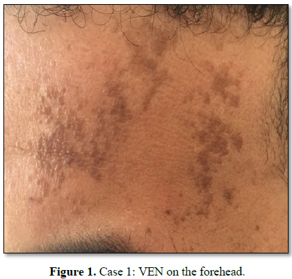
30 year old man presented with multiples papules on his forehead that he had had since he was thirteen. On clinical examination, we observed multiples hyper pigmented, keratosis and polycyclic plaques (Figure 1).
Case 2
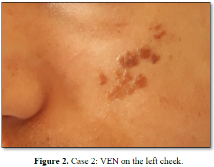
11 year old boy consulted for similar lesions on the left cheek. The lesions had been present since five years. Physical examination finds some brownish papule coalesce to form a serpiginous plaque (Figure 2).
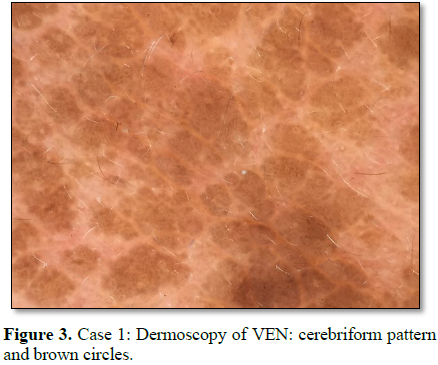
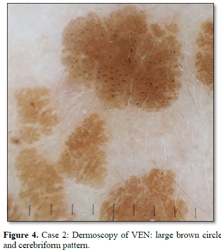
On dermoscopic evaluation, we observed in both cases a cerebriform pattern with pseudo-comedones and nib borde (Figure 3), as well as large brown circles with variable dimensions and are located at the periphery or in the middle of the lesion (Figure 4). The histopathology showed features of verrucous epidermal naevus.
No difference between adult and pediatric appearance was noticed.
DISCUSSION
Various dermoscopic studies of sebaceous hamartomas or seborrheic keratosis have been published. However, few dermoscopic descriptions of VEN had already been made [2].
According to Canning, epidermal nevi present dermoscopically a cerebriform pattern, more often associated with seborrheic keratosis. Carbotti [2] has described, as we do, brown circles that may correlate histologically with the characteristic arrangement of pigmented keratinocytes surrounding the dermal papillae and which may be considered a characteristic feature of VENs.
CONCLUSION
Cerebriform pattern and large brown circles represent a specific dermoscopic feature for the diagnosis of verrucous epidermal nevus.
1.
Lozano B (2017) Dermoscopic features in three cases
of rounded and velvety epidermal naevus. Australas J Dermatol 58: 325-326.
2.
Carbotti M (2016) Dermoscopy of verrucous epidermal
nevus: Large brown circles as a novel feature for diagnosis. Int J Dermatol 55:
653-656.
QUICK LINKS
- SUBMIT MANUSCRIPT
- RECOMMEND THE JOURNAL
-
SUBSCRIBE FOR ALERTS
RELATED JOURNALS
- International Journal of AIDS (ISSN: 2644-3023)
- Stem Cell Research and Therapeutics (ISSN:2474-4646)
- Journal of Immunology Research and Therapy (ISSN:2472-727X)
- International Journal of Clinical Case Studies and Reports (ISSN:2641-5771)
- Oncology Clinics and Research (ISSN: 2643-055X)
- International Journal of Surgery and Invasive Procedures (ISSN:2640-0820)
- Journal of Spine Diseases


