866
Views & Citations10
Likes & Shares
The study objective was to investigate the effect of the Consciousness Energy Treated test formulation on vital organs like bones, heart, liver, lungs and brain using various cell-based assays. The test formulation and the cell media was divided into two parts; one untreated (UT) and other part received the Biofield Energy Treatment remotely by a renowned Biofield Energy Healer, Shirley Theresa Holmlund, Canada and was labeled as the Biofield Energy Treated (BT) test formulation/media. Cell viability data suggested that the test formulation was safe and non-toxic in six different cells. The Biofield Energy Treated Medium (BT-Med) + Biofield Treated Test Item (BT-TI) group showed 115.6% and 53.3% restoration of viable cells at 10 and 25 µg/mL, respectively in human cardiac fibroblasts cells (HCF) compared to the UT-Med + UT-TI group. Moreover, the BT-Med + UT-TI group showed 113.5% and 73.5% restoration of cell viability at 0.1 and 1 µg/mL, respectively in human hepatoma cells (HepG2) compared to the untreated group. Furthermore, 101.1%, 829.8% and 698.9% restoration of cell viability was observed in adenocarcinomic human alveolar basal epithelial cells (A549) by UT-Med + BT-TI, BT-Med + UT-TI and BT-Med + BT-TI groups, respectively at 10 µg/mL compared to the untreated. The alkaline phosphatase (ALP) level was significantly increased by 97.9% and 69.7% in the UT-Med + BT-TI and BT-Med + BT-TI groups, respectively at 50 µg/mL in human bone osteosarcoma cells (MG-63) compared to the untreated. Additionally, the level of ALP was significantly increased by 58.2% in the BT-Med + BT-TI group at 1 µg/mL in human endometrial adenocarcinoma cells (Ishikawa) compared to the untreated. The percent protection of HCF (heart) cells (decreased of LDH activity) was significantly increased by 67.4% (at 0.1 µg/mL), 80.4% (at 0.1 µg/mL) and 119.8% (at 10 µg/mL) in the UT-Med + BT-TI, BT-Med + UT-TI, BT-Med + BT-TI groups, respectively compared to the untreated. The percent protection of HepG2 (liver) cells (decreased of ALT activity) was significantly increased by 57.6% and 82.5% in the BT-Med + UT-TI group at 25 and 63 µg/mL, respectively; while 123.9% at 10 µg/mL in the BT-Med + BT-TI group compared to untreated. The percent protection of A549 (lungs) cells (increased of SOD activity) was significantly increased by 53.6% and 59% in the UT-Med + BT-TI and BT-Med + BT-TI groups, respectively at 10 µg/mL compared to the untreated. Serotonin level was significantly increased by 85.3% in the UT-Med + BT-TI and BT-Med + BT-TI groups at 0.1 µg/mL as compared to untreated in human neuroblastoma cells (SH-SY5Y). The relative quantification (RQ) of vitamin D receptor (VDR) was significantly increased by 245.9% and 211.5% at 10 and 50 µg/mL, respectively in the UT-Med + BT-TI group; while 174.3% (at 10 µg/mL) in the BT-Med + UT-TI group as compared to the untreated in MG-63 cells. Overall, these results suggest that Biofield Energy Treated test formulation has significantly improved the bones, heart, liver, lungs and brain functional enzymes biomarkers. Altogether data suggest that the Biofield Energy Treatment (The Trivedi Effect®) can be useful to protect and maintain the normal function of each vital organ such as lungs, liver, heart, brain, and bones. Therefore, The Trivedi Effect® can be used as a complementary and alternative therapy against several disorders such as heart attack, coronary artery disease, heart failure, arrhythmias, congenital heart disease, cardiomyopathy, Wilson disease,
Keywords: Biofield energy treatment, The Trivedi
effect®, Cardiac health, Bone health, Liver health, VDR receptor,
Lungs health, Brain health
INTRODUCTION
Bones, heart, liver, lungs and brain
disorders are the major concern of human overall health across the globe. The
World Health Organization (WHO) estimates, in 2016, ~17.5 million people die
due to cardiovascular (heart) disorders, ~3.5 million people die due to lungs
disorders, ~1.3 million people die due to liver disorders around the globe each
year [1]. Moreover, ~1.2 million people most frequently diagnosed adult-onset
brain disorders in each year in the USA. [2]. Three main criteria to keep a
healthy heart include the opening blood vessels, strengthening the heart muscle
and controlling free radical damage by antioxidants [3]. The release of liver mitochondrial
enzymes is considered strong evidence for hepatic (liver) necrosis, which is
associated with an increased production of reactive oxygen species (ROS) that
leads to hepatic lipid peroxidation [4-6]. Oxidative stress in the respiratory
system increases the production of mediators of pulmonary inflammation and
initiate or promote mechanisms of carcinogenesis [7]. The lung is one of the
major organs, which is highly exposed by various oxidants, i.e., endogenous and
exogenous oxidants (cigarette smoke, mineral dust, ozone and radiation). These
oxidants produce free radicals, while reactive oxygen species (ROS) and
reactive nitrogen species (RNS) are produced by phagocytes as well as by
alveolar, polymorph nuclear, bronchial and different endothelial cells [8].
However, the role of oxidative stress in the pathogenesis of lung diseases has
been widely reported such as asthma, chronic obstructive pulmonary disease
(COPD), lung malignancies and parenchymal lung diseases like idiopathic
pulmonary fibrosis and lung granulomatous diseases [9]. Serotonin
(5-hydroxytryptamine, 5-HT) is among the brain’s neuromodulators responsible
for behavior and understanding [10]. Apart from medicines, non-pharmacologic
methods that can increase serotonin by increasing recognition and happiness and
well-being. These factors can protect against mental and physical disorders
[11]. There is currently no universally accepted test formulation, which
improve the organ health biomarkers. With this respect, the novel test
formulation was designed on the basis of best scientific literature, which is
the combination of herbal products viz.
Panax ginseng extract and beta
carotene, minerals viz. calcium
chloride, magnesium gluconate, zinc chloride, sodium selenate, ferrous sulfate,
and vitamins viz. vitamin B12,
vitamin D3, ascorbic acid and vitamin B6. This formulation is
designed for overall functioning of the organs that can results in improved
overall health conditions against many pathological conditions such as lung
disorder, liver disorder, breast cancer, liver cancer, aging, muscle damage,
and overall health. Minerals and vitamins present in the test formulation
provide significant functional support to all the vital organs [12-14]. In
addition, Panax ginseng is one of the
best reported medicinal plants that improve mental, physical abilities,
cognitive health and is potent immuno modulator [15,16].
Various study data suggested the effect of
Energy Therapy in cancer patients through therapeutic touch [17]; massage
therapy [18], etc. Complementary and Alternative Medicine (CAM) therapies are
preferred model of treatment, among which Biofield Therapy (or Healing
Modalities) is one approach to enhance emotional, mental, physical and human
wellness. The National Center of Complementary and Integrative Health (NCCIH)
has recognized and allowed Biofield Energy Healing as a CAM approach in
addition to other therapies and medicines such as natural products,
chiropractic/osteopathic manipulation, Qi Gong, deep breathing, Tai Chi, yoga,
meditation, massage, special diets, healing touch, relaxation techniques,
traditional Chinese herbs and medicines, naturopathy, movement therapy,
homeopathy, progressive relaxation, guided imagery, pilates, acupuncture,
acupressure, Reiki, rolfing structural integration, hypnotherapy, Ayurvedic
medicine, mindfulness, essential oils, aromatherapy, and cranial sacral
therapy. The Human Biofield Energy has subtle energy that has the capacity to
work in an effective manner [19]. CAM therapies have been practiced worldwide
with reported clinical benefits in different health disease profiles [20]. This
energy can be harnessed and transmitted by the practitioners into living and
non-living things via the process of Biofield Energy Healing. The Biofield
Energy Treatment, the Trivedi Effect®, has been reported to have a
significant impact in the field of cancer research [21,22], materials science
[23,24], microbiology [25,26], agriculture [27,28], nutraceuticals [29,30] and
biotechnology [31,32]. Further, the Trivedi Effect® also
significantly improved bioavailability of various low bioavailable compounds
[33-35], an improved overall skin health [36,37], bone health [38-40], human
health and wellness. Based on the excellent outcomes of the Biofield Energy
Therapy in wide spectrum of areas, the authors intend to see the impact of the
Biofield Energy Healing Treated test formulation on the function of vital
organs such as bones, heart, liver, lungs and brain specific biomarkers in
different cell-lines.
MATERIALS AND METHODS
Chemicals and
reagents
Ferrous sulfate, vitamin B6, vitamin D3,
vitamin B12, calcium chloride, naringenin, trimetazidine (TMZ),
3-(4,5-Dimethylthiazol-2-yl)-2,5-Diphenyltetrazolium Bromide (MTT) and
ethylenediaminetetraacetic acid (EDTA) were obtained from Sigma Chemical Co.
(St. Louis, MO). Zinc chloride, magnesium gluconate, β-carotene and calcitriol
were purchased from TCI chemicals, Japan. Panax
ginseng extract obtained from Panacea Phytoextracts, India. Sodium selenate
and ascorbic acid were obtained from Alfa Aesar, India. Silymarin and curcumin
were obtained from Sanat Chemicals, India and quercetin obtained from Clearsynth,
India. Reverse Transcription Kit, RNeasy Mini Kit and Syber Green PCR kits were
procured from Quagen, India. All the other chemicals used in this experiment
were analytical grade procured from India.
Biofield energy
healing strategy
The test formulation was the combination of
eleven ingredients viz. calcium
chloride, Panax ginseng extract,
vitamin B12, β-carotene, vitamin D3, zinc chloride, magnesium
gluconate, sodium selenate, ferrous sulfate, ascorbic acid and vitamin B6. The
test formulation and the cell media was divided into two parts; one untreated
(UT) and other part received the Biofield Energy Treatment remotely by a
renowned Biofield Energy Healer, Shirley Theresa Holmlund, under laboratory
conditions for ~3 min through healer’s unique Biofield Energy Transmission
process and were labeled as the Biofield Energy Treated (BT) test
formulation/media. Further, the untreated group was treated with a “sham”
healer for comparison purposes. The “sham” healer did not have any knowledge
about the Biofield Energy Healing Treatment. The Biofield Energy Healer was
located in the Canada; however the test items were located in the research
laboratory of Dabur Research Foundation, New Delhi, India. Biofield Energy
Healer did not visit the laboratory, nor had any contact with the test samples.
After that, the Biofield Energy Treated and untreated test items were kept in
similar sealed conditions and used for the study as per the study plan.
Assessment of cell
viability using MTT assay
Cells were counted using hemocytometer and
plated in 96-well plates at the specific density described in Table 1. The cells were then incubated
overnight under growth conditions to allow cell recovery and exponential
growth. Following overnight incubation, cells were treated with different
concentrations of test formulations (BT/UT). Following respective treatments,
cells were incubated in a CO2 incubator at 37°C, 5% CO2
and 95% humidity and incubated for time period mentioned in Table 1. After incubation, the plates
were taken out and 20 µL of 5 mg/mL of MTT 3-(4,5-dimethylthiazol-2-yl)-2,5-diphenyl
tetrazolium bromide solution was added to all the wells followed by additional
incubation for 3 h at 37°C. The supernatant was aspirated and 150 µL of DMSO
was added to each well to dissolve formazan crystals. The absorbance of each
well was read at 540 nm using Synergy HT microplate reader. The percentage
cytotoxicity at each tested concentration of TI was calculated using Equation
1:
% Cytotoxicity = [(R-X)/R] *100............ (1)
Where, X=Absorbance of treated cells;
R=Absorbance of untreated cells
The concentrations exhibiting percentage
cytotoxicity <30% was considered as non-cytotoxic [41].
Evaluation of the cytoprotective effect of
the formulation
Cells (human cardiac fibroblasts-HCF; human
hepatoma cells-HepG2; and adenocarcinomic human alveolar basal epithelial
cells-A549) were counted and plated in suitable medium followed by overnight
incubation. The cells were then treated with the test items/positive control at
the non-cytotoxic concentrations for 24 h. After 24 h, oxidative stress was
given to the cells using 10 mM t-BHP
for 3.5 h. The untreated cells served as a control that did not receive any
treatment and was maintained in cell growth medium only. Cells treated with 10
mM of t-BHP alone served as negative
control. After 3.5 h of incubation with t-BHP
the above plates were taken out and cell viability was determined by MTT assay.
The percentage protection corresponding to each treatment was calculated using
Equation 2:
% Protection = [(Absorbancesample-Absorbancet-BHP)]*100
/ [Absorbanceuntreated-Absorbancet_BHP]...............
(2)
Assessment of
alkaline phosphatase (ALP) activity
The cells (human bone osteosarcoma cells-MG-63
and human endometrial adenocarcinoma cells-Ishikawa) were counted using a
hemocytometer and plated in 24-well plates at the density corresponding to 1 ×
104 cells/well in phenol-free DMEM supplemented with 10% CD-FBS.
Following the respective treatments, the cells in the above plate were
incubated for 48 h in CO2 incubator at 37°C, 5% CO2 and
95% humidity. After 48 h of incubation, the plates were taken out and processed
for the measurement of ALP enzyme activity. The cells were washed with 1x PBS
and lysed by freeze-thaw method, i.e., incubation at -80°C for 20 min followed
by incubation at 37°C for 10 min. To the lysed cells, 50 µL of substrate
solution, i.e., 5 mm of p-nitrophenyl
phosphate (pNPP) in 1 M
diethanolamine and 0.24 mm magnesium chloride (MgCl2) solution (pH
10.4) was added to all the wells followed by incubation for 1 h at 37°C. The
absorbance of the above solution was read at 405 nm using Synergy HT microplate
reader (Biotek, USA). The absorbance values obtained were normalized with
substrate blank (pNPP solution alone)
absorbance values. The percentage increase in ALP enzyme activity with respect
to the untreated cells (baseline group) was calculated using Equation 3:
% Increase in ALP = {(X-R)/R}*100 (3)
Where, X=Absorbance of cells corresponding to
positive control and test groups; R=Absorbance of cells corresponding to
baseline group (untreated cells)
Estimation of
lactate dehydrogenase (LDH) in human cardiac fibroblasts (HCF)
The human cardiac fibroblasts (HCF) Cells
were counted and plated at the density of 0.25 × 10⁶ cells/ well in
24-well plates in cardiac fibroblast specific medium followed by overnight
incubation. The cells were then treated with the test formulation/positive
control at the non-cytotoxic concentrations for 24 h. After 24 h, oxidative
stress was given to the cells using 10 mM t-BHP
for 3.5 h. The untreated cells were served as control that did not receive any
treatment and were maintained in cell growth medium only. Cells treated with 10
mM of t-BHP alone served as the
negative control. After 3.5 h of incubation with t-BHP the above plates were taken out and LDH activity was
determined using LDH activity kit as per manufacturer’s instructions. The
percent increase in LDH activity was calculated using Equation 4:
% Increase = [(LDH activitysample-LDH
activityt-BHP)]*100 / [LDH activityuntreated-LDH activityt_BHP]..............
(4)
Estimation of ALT in liver
cells (HepG2)
The human
hepatoma cells (HepG2) were counted and plated at the density of 5 × 10⁴
cells/well in 48-well plates in DMEM media followed by overnight incubation. The cells were then treated with the test formulation/positive
control at the non-cytotoxic concentrations for 24 h. After 24 h, oxidative
stress was given to the cells using 400 µM t-BHP
for 3.5 h. The untreated cells served as control that did not receive any
treatment and were maintained in cell growth medium only. Cells treated with
400 µM of t-BHP alone served as
negative control. After 3.5 h of incubation with t-BHP the above plates were taken out and ALT activity was
determined using ALT activity kit as per manufacturer’s instructions. The
percent increase in ALT activity was calculated using Equation 5:
% Increase = [(ALT activitysample-ALT
activityt-BHP)]*100/ [ALT activityuntreated-ALT activityt_BHP].............
(5)
Estimation of
superoxide dismutase (SOD) in lung (A549) cells
The adenocarcinomic human alveolar basal
epithelial cells (A549) were counted and plated at the density of 1 × 10⁴
cells/well in 24-well plates in DMEM followed by overnight incubation. The
cells were then treated with the test formulation/positive control at the
non-cytotoxic concentrations along with 100 µM t-BHP to induce oxidative stress. The untreated cells served as
control that did not receive any treatment and were maintained in cell growth
medium only. Cells treated with 100 µM of t-BHP
alone served as negative control. After 24 h of incubation with t-BHP the above plates were taken out
and SOD activity was determined using SOD activity kit as per manufacturer’s
instructions. The percent increase in SOD activity was calculated using
Equation 6:
%
Increase in SOD activity
= ((X-R)/R)*100................ (6)
Where, X=SOD activity corresponding to test
item or positive control; R=SOD activity corresponding to control group
Estimation of
serotonin in neuronal cells (SH-SY5Y)
The human neuroblastoma (SH-SY5Y) cells were
counted and plated at the density of 10 × 104 cells/well in 96-well
plates followed by overnight incubation. The cells were then treated with the
test items/positive control at the non-cytotoxic concentrations. The untreated
cells served as control that did not receive any treatment and were maintained
in cell growth medium only. The treated cells were incubated for 24 h.
Serotonin release was determined by ELISA as per manufacturer’s protocol. The
percent increase in serotonin levels was calculated using Equation 7:
[(X-R)/R]*100................ (7)
Where, X=Serotonin levels corresponding to
test item or positive control; R=Serotonin levels corresponding to control
group
Effect of test formulation on vitamin D receptor (VDR) in bone
(MG-63) cells
The human bone
osteosarcoma (MG-63) cells were counted using the hemocytometer were plated at a density of 2 × 10⁵ cells/well in 6-well plates followed by overnight incubation. The cells were then sera starved for 24 h and treated with the test
formulation/positive control at the non-cytotoxic concentrations. The untreated cells that served as
control that did not receive any treatment and were maintained in cell growth
medium only. The treated cells were incubated for 24 h and VDR expression was determined by Q-PCR using VDR specific primers.
Cells were harvested by scrapping and washed with PBS. Cell pellets obtained
were analyzed for VDR gene expression using human VDR specific primers:
Forward: 5’-GCTGACCTGGTCAGTTACAGCA-3’, Reverse: 5’-CACGTCACTGACGCGGTACTT-3’. VDR gene
expression was normalized using House-keeping (HK) reference. Relative
quantification (RQ) of VDR gene in Biofield Energy Treated cells was calculated
with respect to the untreated cells using Equation 8:
RQ = 2-N................ (8)
Where N is the relative Threshold Cycle (CT)
value of treated sample with respect to the untreated sample
STATISTICAL ANALYSIS
All the values were represented as Mean ± SD
(standard deviation) of three independent experiments. The statistical analysis
was performed using SigmaPlot statistical software (v11.0). For two groups
comparison student’s t-test was used.
For multiple group comparison, one-way analysis of variance (ANOVA) was used
followed by post-hoc analysis by Dunnett’s test. Statistically significant
values were set at the level of p ≤
0.05.
RESULTS AND
DISCUSSION
Cell viability using
MTT assay
Determination of non-cytotoxic concentration
of the formulation and positive controls by MTT cell viability assay was used
in terms of percent viable cells in six (6) different cell-lines viz. MG-63, Ishikawa, A549, HepG2, HCF
and SH-SY5Y. Based on the percent cell viability data, it was observed that the
formulation and positive controls were safe and non-toxic at the tested
concentrations in six different cell lines and selected for other parameters
analysis.
Evaluation of
cytoprotective effect of the test formulation
The cytoprotective activity of the novel
proprietary test formulation on vital organs like liver, heart and lungs was
examined in in vitro cell-based assay
under the stimulation of tert-butyl
hydroperoxide (t-BHP) induced
oxidative stress. t-BHP has been
routinely used for the induction of oxidative stress in various cells [42]. The
cytoprotective activity of the test formulation on the restoration of cell
viability was determined against t-BHP
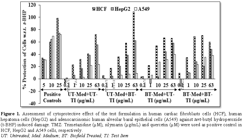
induced cell damage and the result is shown in Figure 1. Trimetazidine (TMZ) was used as positive control in human
cardiac fibroblasts cells (HCF) and showed, restoration of cell viability by
34.01%, 60.04% and 98.31% at 5, 10 and 25 µg/mL, respectively compared to the t-BHP induced group. Besides, the test
formulation showed 52.7% restoration of cell viability at 1 µg/mL in the BT-Med
+ BT-TI group as compared to the UT-Med + UT-TI group. Moreover, at 10 µg/mL
the UT-Med + BT-TI, BT-Med + UT-TI and BT-Med + BT-TI groups showed 11.6%,
67.5% and 115.6% restoration of cell viability, respectively than UT-Med +
UT-TI group. Additionally, the test formulation showed 41.9%, 62% and 71.8%
restoration of cell viability at 25 µg/mL in the UT-Med + BT-TI, BT-Med + UT-TI
and BT-Med + BT-TI groups, respectively as compared to the UT-Med + UT-TI
group. Further, at 63 µg/mL the test formulation showed 51.2% restoration of
cell viability in the UT-Med + BT-TI group than UT-Med + UT-TI group (Figure 1). Silymarin was used as
positive control in human hepatoma cells (HepG2) resulted, restoration of cell
viability by 31.63%, 64.63% and 74.64% at 5, 10 and 25 µg/mL, respectively
compared to the t-BHP induced group. The test formulation showed 113.5%
restoration of cell viability at 0.1 µg/mL in the BT-Med + UT-TI group as
compared to the UT-Med + UT-TI group. Moreover, at 1 µg/mL the BT-Med + UT-TI
group showed 73.5% restoration of cell viability than UT-Med + UT-TI group. The
test formulation showed 20.7% and 66.3% restoration of cell viability at 10
µg/mL in the UT-Med + BT-TI and BT-Med + BT-TI groups, respectively as compared
to the UT-Med + UT-TI group (Figure 1).
Quercetin was used as positive control in adenocarcinomic human alveolar basal
epithelial cells (A549) resulted, restoration of cell viability by 30.8%,
69.33% and 72.22% at 5, 10 and 25 µg/mL, respectively compared to the t-BHP
induced group. Besides, the test formulation showed 60% restoration of cell
viability at 1 µg/mL in the UT-Med + BT-TI group compared to the UT-Med + UT-TI
group. Moreover, at 10 µg/mL the UT-Med + BT-TI, BT-Med + UT-TI and BT-Med +
BT-TI groups showed 101.1%, 829.8% and 698.9% restoration of cell viability,
respectively than UT-Med + UT-TI group. Additionally, the test formulation
showed 46.2% and 63.3% restoration of cell viability at 25 µg/mL in the BT-Med
+ UT-TI and BT-Med + BT-TI groups, respectively as compared to the UT-Med +
UT-TI group. Further, the test formulation showed 67.6% and 62.8% restoration
of cell viability at 63 µg/mL in the BT-Med + UT-TI and BT-Med + BT-TI groups,
respectively compared to the UT-Med + UT-TI group (Figure 1). Oxidative stress is linked with a wide variety of
inflammatory and metabolic disease conditions. Besides, cumulative damage of
cells by free radicals inadequately neutralized by antioxidants [43]. The study
results suggest that Biofield Treatment has significantly protects t-BHP induced cardiotoxicity,
hepatotoxicity and lung cell toxicity which could be due to The Trivedi Effect®-Biofield
Energy Healing as free radical scavenging activity. Therefore, Biofield Energy
Healing Treatment could be used for the management of cardiovascular, liver and
various lung disorders.
Assessment of
alkaline phosphatase (ALP) activity
The effect of the test formulation on
bone-specific alkaline phosphatase level is shown in Figure 2. The positive control, calcitriol showed 20.03%, 22.71%
and 36.75% increase the level of ALP at 0.1, 1 and 10 nM, respectively in MG-63
cells. The UT-Med + BT-TI group showed 44.9% increase the level of ALP in with
respect to the UT-Med + UT-TI group at 1 µg/mL. At 10 µg/mL, the percent ALP
was significantly increased by 47.8%, 12.4% and 48.1% in the UT-Med + BT-TI,
BT-Med + UT-TI and BT-Med + BT-TI groups, respectively compared to the UT-Med +
UT-TI group. Further, the percent ALP was significantly increased by 97.9%,
13.7% and 69.7% in the UT-Med + BT-TI, BT-Med + UT-TI and BT-Med + BT-TI
groups, respectively at 50 µg/mL compared to the UT-Med + UT-TI group (Figure 2). Besides, the positive
control, naringenin showed 25.93%, 49.23% and 151.85% increase the level of ALP
at 0.1, 1 and 10 nM, respectively in Ishikawa cells. ALP percent was
significantly increased by 29%, 19% and 58.2% in the UT-Med + BT-TI, BT-Med +
UT-TI, BT-Med + BT-TI groups, respectively compared to the UT-Med + UT-TI group
at 50 µg/mL (Figure 2). Numerous
experimental data reported that lower level of serum alkaline phosphatase (ALP)
can improve the bone mineral density (BMD) [43]. Thus, for the detection of
bone specific biochemical marker in serum can be clinically useful in evaluating
the progress of the bone healing process [44]. In this experiment, the level of
ALP was revealed that the Biofield Energy Healing Treated test formulation
significantly increased the level of ALP expression, which might be very
helpful to the patients suffering from various bone-related disorders.
Estimation of
lactate dehydrogenase (LDH) activity in human cardiac fibroblasts (HCF)
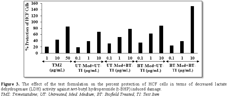
The effect of the test items on the percent
protection of HCF cells in terms of decreased level of lactate dehydrogenase
(LDH) activity is shown in Figure 3.
The positive control, trimetazidine (TMZ) exhibited 20.53%, 43.08% and 85.86%
protection of HCF cells (decreased of LDH activity) compared to the t-BHP group. The percent protection of
HCF cells (decreased of LDH activity) was significantly increased by 67.4%,
80.4% and 28.9% in the UT-Med + BT-TI, BT-Med + UT-TI and BT-Med + BT-TI
groups, respectively at 0.1 µg/mL as compared to the UT-Med + UT-TI group.
Moreover, at 1 µg/mL, the percent protection of HCF cells (decreased of LDH
activity) was significantly increased by 36.4% and 65.6% in the UT-Med + BT-TI
and BT-Med + UT-TI groups, respectively as compared to the UT-Med + UT-TI
group. Further, percent protection of HCF cells (decreased of LDH activity) was
also significantly increased by 13.8%, 28.2%, and 119.8% in the UT-Med + BT-TI,
BT-Med + UT-TI and BT-Med + BT-TI groups, respectively at 10 µg/mL as compared
to the UT-Med + UT-TI group (Figure 3).
The lactate dehydrogenase (LDH) isoenzymes in serum was used for the late
diagnosis of myocardial infarction [45], prognosis and management of certain
tumors [46]. The study results found that there was a significant reduction of
LDH level after Biofield Energy Treatment and protect heart cells, which might
be helpful to resist against various pathological conditions like tissue
injury, necrosis, hemolysis or malignancies, hypoxia, etc. It also indicating
that the heart cells acted normally under stress and anaerobic condition and
improved overall heart function.
Estimation of alanine amino
transferase (ALT) activity in HepG2 cells
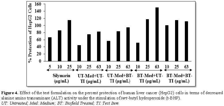
The effect of
the test formulation on protection of HepG2 cells in terms of decrease alanine
amino transferase (ALT) activity is shown in Figure 4. The positive control, silymarin exhibited 66.35%, 85.83%
and 114.38% protection of HepG2 cells (decreased of ALT activity) at 5, 10 and
25 µg/mL, respectively as compared to the tert-butyl
hydroperoxide (t-BHP). The protection
of HepG2 cells (decreased of ALT activity) was significantly increased by
26.6%, 15% and 123.9% at 10 µg/mL in the UT-Med + BT-TI, BT-Med + UT-TI and
BT-Med + BT-TI groups, respectively as compared to the UT-Med + UT-TI group.
Moreover, at 25 µg/mL percent protection of HepG2 cells (decreased of ALT
activity) was significantly increased by 11.3%, 57.6% and 54% in the UT-Med +
BT-TI, BT-Med + UT-TI and BT-Med + BT-TI groups, respectively as compared to
the UT-Med + UT-TI group. Further, protection of HepG2 cells (decreased of ALT
activity) was also significantly increased by 15.3%, 82.5% and 35% in the
UT-Med + BT-TI, BT-Med + UT-TI and BT-Med + BT-TI groups, respectively at 63
µg/mL as compared to the UT-Med + UT-TI group (Figure 4). Abnormal levels of liver enzyme like ALT cause liver
damage or change in bile flow capacity by either accompanying biochemical
picture in a patient with symptoms or signs [47]. This enzyme can catalyze the
reversible transformation of α-ketoacids into amino acids and play as a
predictor of mortality independent of liver disease [48]. Here, the Biofield
Energy Treatment significantly protect liver hepatocytes in terms of reducing
the level of transaminases enzyme, ALT compared to the t-BHP inducing group, which
might be due to Consciousness Energy Healing Treatment to the test formulation.
Estimation of superoxide dismutase (SOD) activity in
adenocarcinomic human alveolar basal epithelial cells (A549)
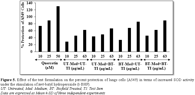
The effect of
the test formulation on the protection of lungs cells (A549) in terms of
increased super oxide dismutase (SOD) activity is shown in Figure 5. The positive control, showed 74.04%, 89.75% and 129.89%
protection of A549 (lungs) cells (increased of SOD activity) at 10, 25 and 63
µM, respectively compared to the t-BHP
group. The percent protection of A549 (lungs) cells (increased of SOD activity)
was significantly increased by 53.6%, 17.4% and 59% at 10 µg/mL in the UT-Med +
BT-TI, BT-Med + UT-TI and BT-Med + BT-TI groups, respectively compared to the
UT-Med + UT-TI group. Moreover, at 63 µg/mL, the percent protection of A549
(lungs) cells (increased of SOD activity) was significantly increased by 6.7%,
38.1% and 44.1% in the UT-Med + BT-TI, BT-Med + UT-TI and BT-Med + BT-TI
groups, respectively as compared to the UT-Med + UT-TI group (Figure 5). Cells contain a large
number of antioxidants like superoxide dismutase (SOD), catalase, glutathione
peroxidase etc. to prevent or repair the damage of cells caused by ROS, as well
as to regulate redox-sensitive signaling pathways [49]. Another, researcher
reported that pulmonary superoxide dismutase (SOD) activity become more active
in aged peoples and plays an important role against various respiratory
disorders such as asthma, chronic obstructive pulmonary disease (COPD), lung
malignancies, etc. [50]. Data found a significant increased SOD level after
Biofield Energy Treatment in A549 cells, which might be helpful to resist
against various pathological conditions like oxidative stress and related
adverse effect. It also indicating that the lung cells acted normally and
improved overall respiratory activities.
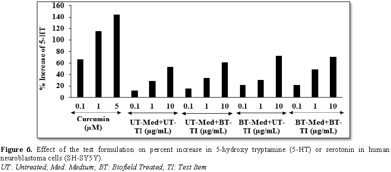
Effect of test formulation on serotonin in human neuroblastoma (SH-SY5Y) cells
The effect of test formulation on serotonin
level is shown in Figure 6. The positive control
showed 66.33%, 115.13% and 143.41% increase the level of serotonin. The level
of serotonin was significantly increased by 33.3%, 85.3% and 85.3% in the
UT-Med + BT-TI, BT-Med + UT-TI, BT-Med + BT-TI groups, respectively at 0.1
µg/mL compared to the UT-Med + UT-TI group. Moreover, at 1 µg/mL, 5-HT level
was significantly increased by 21.9%, 7.7% and 74.2% in the UT-Med + BT-TI,
BT-Med + UT-TI and BT-Med + BT-TI groups, respectively as compared to the
UT-Med + UT-TI group. Further, the serotonin level was significantly increased
by 13.8%, 35.8% and 33.6% in the UT-Med + BT-TI, BT-Med + UT-TI and BT-Med +
BT-TI groups, respectively at 10 µg/mL as compared to the UT-Med + UT-TI group (Figure 6). Serotonin (5-HT) is a
neurotransmitter responsible for stress, anxiety, aggressive behavior, and many
more [51]. Recent studies reported that brain endothelium is the specific
target for serotonin and actively involved in the regulation of the blood-brain
barrier (BBB) permeability and the cerebral blood flow via receptor-mediated mechanisms [52]. Thus, this experimental data
suggested that Biofield Energy Healing Treated novel formulation significantly
improved the serotonin level, which would be highly useful against various
neurodegenerative diseases and other age-related disorders and improved the
normal functioning of the brain tissues.
UT: Untreated; Med:
Medium; BT: Biofield Treated; TI: Test Item
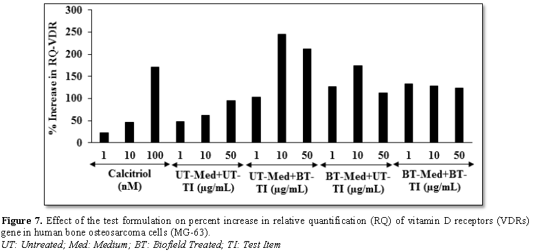
Effect of
test formulation on vitamin D receptors (VDRs)
Human bone osteosarcoma cells (MG-63) were
treated with the test formulation and the effect on vitamin D receptor (VDR)
expression was determined using quantitative-polymerase chain reaction (Q-PCR)
amplification. VDR-relative threshold cycle (VDR-CT) values were obtained from
PCR amplification. Relative quantification (RQ) of VDR was calculated from the
VDR-CT and house-keeping (HK)-CT values for MG-63 cells treated with test
formulation and positive control is shown in Figure 7. The positive
control (calcitriol) showed 22.26%, 46.41% and 171.32% increase of RQ of VDR at
1, 10 and 100 NM, respectively. Moreover, RQ-VDR expression was significantly
increased by 103.5%, 126.9% and 133% in the UT-Med +
BT-TI, BT-Med + UT-TI and BT-Med + BT-TI groups, respectively at 1 µg/mL
compared to the UT-Med + UT-TI group. Additionally, at 10 µg/mL the VDR level
was significantly increased by 245.9%, 174.3% and 128.7% in the UT-Med + BT-TI,
BT-Med + UT-TI, and BT-Med + BT-TI groups, respectively compared to the UT-Med
+ UT-TI group. Further, VDR level was also significantly increased by 211.5%,
112.7% and 123.9% in the UT-Med + BT-TI, BT-Med + UT-TI and BT-Med + BT-TI
groups, respectively at 50 µg/mL compared to the UT-Med + UT-TI group. Vitamin
D can enhanced calcium absorption in the small intestine and stimulates
production of active metabolites 1, 25[OH]2 D3 in the
kidney [53]. The hormone then interacts with the vitamin D receptor (VDR) in
intestinal cells and complexes with the retinoic acid x receptor (RXR) in the
nucleus [54]. This complex binds to the vitamin-D-responsive element (VDRE) of
the calcium channel which increases uptake of calcium into the cells and
increases the absorption of calcium [55]. Overall, the Consciousness Energy
Treated test formulation has excellently increased the expression of VDRs,
which might be helpful to bind more active vitamin D3 metabolites
and that ultimately can improve the more physiological functions of vitamin D
and simultaneously improved bone cell growth and development.
CONCLUSION
The study findings showed that the tested
novel test formulation was safe and non-toxic based on the MTT cell viability
assay in six tested cells. The treatment group like BT-Med + BT-TI showed
115.6% restoration of cell viability at 10 µg/mL in human cardiac fibroblasts
cells (HCF) compared to the UT-Med + UT-TI group. Moreover, the BT-Med + UT-TI
group showed 113.5% and 73.5% restoration of cell viability at 0.1 and 1 µg/mL, respectively in human
hepatoma cells (HepG2) compared to the untreated group. Additionally, 101.1%,
829.8%, and 698.9% restoration of cell viability at 10 µg/mL in adenocarcinomic
human alveolar basal epithelial cells (A549) compared to the untreated group.
Alkaline phosphatase (ALP) activity was significantly increased by 97.9%
and 69.7% in the UT-Med + BT TI and BT-Med + BT TI groups, respectively at 50
µg/mL compared to the untreated in human bone osteosarcoma cells (MG-63).
Moreover, ALP activity was
significantly increased by 58.2% in the BT-Med + BT-TI group at 1 µg/mL
than untreated group. The percent protection of HCF cells (decreased of LDH
activity) was significantly increased by 80.4% (at 0.1 µg/mL) and 119.8% (at 10
µg/mL) in the BT-Med + UT-TI and BT-Med + BT-TI groups, respectively compared
to the untreated group. The percent protection of HepG2 cells (decreased of ALT
activity) was significantly increased by 82.5% (at 63
µg/mL) and 123.9% (at 10 µg/mL) in the BT-Med + UT-TI and BT-Med + BT-TI
groups, respectively compared to the untreated group. The percent protection of
A549 (lungs) cells (increased of SOD activity) was significantly increased by
59% in the BT-Med + BT-TI group at 10 µg/mL compared to the untreated group.
The serotonin level was significantly increased by 85.3% at 0.1 µg/mL in the
UT-Med + BT-TI and BT-Med + BT-TI groups as compared to the untreated group in
human neuroblastoma cells (SH-SY5Y). The relative quantification (RQ) of vitamin D receptors (VDRs) level was
significantly increased by 245.9% and 211.5% at 10 and 50 µg/mL, respectively
in the UT-Med + BT-TI group; while 174.3% (at 10 µg/mL) in the BT-Med + UT-TI
group compared to the untreated group in MG-63 cells. In conclusion, The Biofield Energy Treatment
significantly improved heart, liver, bones, neuronal and lungs functional
enzymes biomarkers and also protected cardiomyocyte, hepatocyte, osteocytes,
pneumocyte and nerve cells from oxidative damage induced by tert-butyl hydroperoxide (t-BHP). Thus, results suggested that
Biofield Energy Treatment can be used as a complementary and alternative treatment for the prevention of various types
of cardiac disorders (peripheral artery disease, high blood pressure,
congenital heart disease, stroke, congestive heart failure, rheumatic heart
disease, carditis, valvular heart disease, thromboembolic disease and venous
thrombosis, etc.), hepatic disorders (cirrhosis, Wilson disease, liver
cancer, hemochromatosis), and lungs
disorders (Asthma, Emphysema, Chronic bronchitis, Pneumonia, Cystic fibrosis). Further,
it can be useful to improve cell-to-cell messaging, normal cell growth and
differentiation, cell cycling and proliferation, neurotransmission, skin
health, hormonal balance, immune and cardiovascular functions. Moreover, it can
also be utilized in organ transplants (i.e.,
liver, kidney, and heart transplants), aging, hormonal imbalance and various
inflammatory and immune-related disease conditions like Alzheimer’s Disease
(AD), Dermatitis, Asthma, Ulcerative Colitis (UC), Hashimoto Thyroiditis, Pernicious Anemia, Sjogren Syndrome, Aplastic
Anemia, Multiple Sclerosis, Hepatitis, Graves’
Disease, Irritable Bowel Syndrome (IBS), Dermatomyositis,
Diabetes, Myasthenia Gravis, Atherosclerosis, Parkinson’s Disease, Systemic, etc., to Lupus Erythematosus (SLE), stress, improve overall health and
Quality of Life.
ACKNOWLEDGEMENT
Authors gratefully acknowledged to Trivedi
Global, Inc., Trivedi Science and Trivedi Master Wellness for their support. In
addition, authors are thankful for the support of Dabur Research Foundation for
conducting this study.
CONFLICT OF
INTEREST
Authors declare no conflict of interest.
1. Global
Burden of Disease Collaborative Network. Global Burden of Disease Study 2016
(GBD) (2016) Results. Seattle, United States: Institute for Health Metrics and
Evaluation (IHME).
2. Pal S
(2018) Incidence and prevalence of major neurologic disorders. US Pharm 43: 24.
3. Rakesh
S, Arunporn I (2017) Herbal supplements or herbs in heart disease:
Herbiceutical formulation, clinical trials, futuristic developments. J Cardiol
Cardiovasc Ther 3: 555603.
4. Contreras-Zentella
ML, Hernández-Muñoz R (2016) Is liver enzyme release really associated with
cell necrosis induced by oxidant stress? Oxid Med Cell Longev 2016: 3529149.
5. Schmidt
E, Schmidt FW (1970) Aspects of enzyme diagnosis. Med Welt 21: 805-816.
6. Frederiks
WM, Vogels IM, Fronik GM (1984) Plasma ornithine carbamyl transferase level as
an indicator of ischaemic injury of rat liver. Cell Biochem Funct 2: 217-220.
7. Boots
AW, Haenen GR, Bast A (2003) Oxidant metabolism in chronic obstructive
pulmonary disease. Eur Respir J 46: 14S-27S.
8. Romieu
I (2005) Nutrition and lung health. Int J Tuberc Lung Dis 9: 362-374.
9. Kelly
FJ (2005) Vitamins and respiratory disease: Antioxidant micronutrients in
pulmonary health and disease. Proc Nutr Soc 64: 510-526.
10. Fischer
AG, Ullsperger M (2017) An update on the role of serotonin and its interplay
with dopamine for reward. Front Hum Neurosci 11: 484.
11. Anonymous
(2006) A sensible 10 year plan for mental health. Lancet 367: 86.
12. Ryan-Harshman
M, Aldoori W (2005) Health benefits of selected minerals. Can Fam Physician 51:
673-675.
13. Rayman
MP (2000) The importance of selenium to human health. Lancet 356: 233-241.
14. Beard
JL, Connor JR (2003) Iron status and neural functioning. Ann Rev Nutr 23: 41-58.
15. Coleman
CI, Hebert JH, Reddy P (2003) The effects of Panax ginseng on quality of life. J Clin Pharm Ther 28: 5-15.
16. Das
L, Bhaumik E, Raychaudhuri U, Chakraborty R (2011) Role of nutraceuticals in
human health. J Food Sci Technol 49: 173-183.
17. Lutgendorf
SK, Mullen-Houser E, Russell D, Degeest K, Jacobson G, et al. (2010)
Preservation of immune function in cervical cancer patients during
chemoradiation using a novel integrative approach. Brain Behav Immun 24:
1231-1240.
18. Ironson
G, Field T, Scafidi F, Hashimoto M, Kumar M, et al. (1996) Massage therapy is
associated with enhancement of the immune system's cytotoxic capacity. Int J
Neurosci.84: 205-217.
19. Jain
S, Hammerschlag R, Mills P, Cohen L, Krieger R, et al. (2015) Clinical studies
of biofield therapies: Summary, methodological challenges and recommendations.
Glob Adv Health Med 4: 58-66.
20. Rubik
B (2002) The biofield hypothesis: Its biophysical basis and role in medicine. J
Altern Complement Med 8: 703-717.
21. Trivedi
MK, Patil S, Shettigar H, Mondal SC, Jana S (2015) The potential impact of
biofield treatment on human brain tumor cells: A time-lapse video microscopy. J
Integr Oncol 4: 141.
22. Trivedi
MK, Patil S, Shettigar H, Gangwar M, Jana S (2015) In vitro evaluation of biofield treatment on cancer biomarkers
involved in endometrial and prostate cancer cell lines. J Cancer Sci Ther 7:
253-257.
23. Trivedi
MK, Tallapragada RM (2008) A transcendental to changing metal powder
characteristics. Met Powder Rep 63: 22-28, 31.
24. Trivedi
MK, Nayak G, Patil S, Tallapragada RM, Latiyal O (2015) Studies of the atomic
and crystalline characteristics of ceramic oxide nano powders after bio field
treatment. Ind Eng Manage 4: 161.
25. Trivedi
MK, Branton A, Trivedi D, Nayak G, Charan S, et al. (2015) Phenotyping and 16S
rDNA analysis after biofield treatment on Citrobacter
braakii: A urinary pathogen. J Clin Med Genomics 3: 129.
26. Trivedi
MK, Patil S, Shettigar H, Mondal SC, Jana S (2015) An impact of biofield
treatment: Antimycobacterial susceptibility potential using BACTEC 460/MGIT-TB
System. Mycobact Dis 5: 189.
27. Trivedi
MK, Branton A, Trivedi D, Nayak G, Mondal SC, et al. (2015) Morphological
characterization, quality, yield and DNA fingerprinting of biofield energy
treated alphonso mango (Mangifera indica
L.). J Food Nutr Sci 3: 245-250.
28. Trivedi
MK, Branton A, Trivedi D, Nayak G, Mondal SC, et al. (2015) Evaluation of
biochemical marker – Glutathione and DNA fingerprinting of biofield energy
treated Oryza sativa. Am J Biosci 3:
243-248.
29. Trivedi
MK, Branton A, Trivedi D, Nayak G, Plikerd WD, et al. (2017) A Systematic study
of the biofield energy healing treatment on physicochemical, thermal,
structural and behavioral properties of magnesium gluconate. Int J Bioorg Chem
2: 135-145.
30. Parulkar
VR, Trivedi MK, Branton A, Trivedi D, Nayak G, et al. (2018) Improved
metabolism of vitamin D3 in human osteoblasts cells after biofield
energy healing treatment. Am J Lab Med 3: 11-19.
31. Trivedi
MK, Patil S, Shettigar H, Bairwa K, Jana S (2015) Phenotypic and biotypic
characterization of Klebsiella oxytoca:
An impact of biofield treatment. J Microbiol Biochem Technol 7: 203-206.
32. Nayak
G, Altekar N (2015) Effect of biofield treatment on plant growth and
adaptation. J Environ Health Sci 1: 1-9.
33. Branton
A, Jana S (2017) The influence of energy of consciousness healing treatment on
low bioavailable resveratrol in male Sprague Dawley rats. Int J Clin Dev Anat
3: 9-15.
34. Branton
A, Jana S (2017) The use of novel and unique biofield energy healing treatment
for the improvement of poorly bioavailable compound, berberine in male Sprague
Dawley rats. Am J Clin Exp Med 5: 138-144.
35. Branton
A, Jana S (2017) Effect of The biofield energy healing treatment on the
pharmacokinetics of 25-hydroxyvitamin D3 [25(OH)D3] in
rats after a single oral dose of vitamin D3. Am J Pharmacol
Phytother 2: 11-18.
36. Parulkar
VR, Trivedi MK, Branton A, Trivedi D, Nayak G, et al. (2017) The use of
consciousness energy healing based herbomineral formulation for skin anti-aging
strategies. J Food Nutr Sci 5: 96-106.
37. Singh
J, Trivedi MK, Branton A, Trivedi D, Nayak G, et al. (2017) Consciousness
energy healing treatment based herbomineral formulation: A safe and effective
approach for skin health. Am J Pharmacol Phytother 2: 1-10.
38. Anagnos
D, Trivedi K, Branton A, Trivedi D, Nayak G, et al. (2018) Influence of
biofield treated vitamin D3 on proliferation, differentiation and
maturation of bone-related parameters in MG-63 cell-line. Int J Biomed Eng Clin
Sci 4: 6-14.
39. Lee
AC, Trivedi K, Branton A, Trivedi D, Nayak G, et al. (2018) The potential
benefits of biofield energy treated vitamin D3 on bone
mineralization in human bone osteosarcoma cells (MG-63). Int J Nutr Food Scis
7: 30-38.
40. Stutheit
ME, Trivedi K, Branton A, Trivedi D, Nayak G, et al. (2018) Biofield energy
treated vitamin D3: Therapeutic implication on bone health using
osteoblasts cells. Am J Life Sci 6: 13-21.
41. Alía
M, Ramos S, Mateos R, Bravo L, Goya L (2005) Response of the antioxidant
defense system to tert-butyl
hydroperoxide and hydrogen peroxide in a human hepatoma cell line (HepG2). J
Biochem Mol Toxicol 19: 119-128.
42. Vargas-Mendoza
N, Madrigal-Santillán E, Morales-González A, Esquivel-Soto J, Esquivel-Chirino
C, et al. (2014) Hepatoprotective effect of silymarin. World J Hepatol 6:
144-149.
43. Noeman
SA, Hamooda HE, Baalash AA (2011) Biochemical study of oxidative stress markers
in the liver, kidney and heart of high fat diet induced obesity in rats.
Diabetol Metab Syndr 3: 17.
44. Kyd
PA, De Vooghtl K, Kerkhoff F, Thomas E, Fairneyl A (1998) Clinical usefulness
of bone alkaline phosphatase in osteoporosis. Ann Clin Biochem 35: 717-725.
45. Randall
DC, Jones DL (1997) Eliminating unnecessary lactate dehydrogenase testing: A
utilization review study and national survey. Arch Intern Med 14: 1441-1444.
46. Munshi
NC, Anderson KC, Bergsagel PL, Shaughnessy J, Palumbo A, et al. (2011)
Consensus recommendations for risk stratification in multiple myeloma: Report
of the International Myeloma Workshop Consensus Panel 2. Blood 117: 4696-4700.
47. Giannini
EG, Testa R, Savarino V (2005) Liver enzyme alteration: A guide for clinicians.
CMAJ. 172: 367-379.
48. Mathiesen
U, Franzen L, Fryden A, Foberg U, Bodemar G (1999) The clinical significance of
slightly to moderately increased liver transaminase values in asymptomatic
patients. Scand J Gastroenterol 34: 85-91.
49. Weydert
CJ, Cullen JJ (2010) Measurement of superoxide dismutase, catalase and
glutathione peroxidase in cultured cells and tissue. Nat Protoc 5: 51-66.
50. Autor
AP, Frank L, Roberts RJ (1976) Developmental characteristics of pulmonary
superoxide dismutase: Relationship to idiopathic respiratory distress syndrome.
Pediatr Res 10: 154-158.
51. Dorszewska
J, Prendecki M, Oczkowska A, Rozycka A, Lianeri M, et al. (2013) Polymorphism
of the COMT, MAO, DAT, NET and 5-HTT genes and biogenic amines in Parkinson’s disease.
Curr Genomics 14: 518-533.
52. Brust
P, Friedrich A, Krizbai IA, Bergmann R, Roux F, et al. (2000) Functional
expression of the serotonin transporter in immortalized rat brain micro vessel
endothelial cells. J Neurochem 74: 1241-1248.
53. Khosla
S (2001) The OPG/RANKL/RANK system. Endocrinology 142: 5050-5055.
54. Bouillon
R (2001) Vitamin D: from photosynthesis, metabolism, and action to clinical
applications. In: De Groot L, Jameson JL, Burger HG, editors. Endocrinology. 3rd
Edn. Saunders; Philadelphia, USA, pp: 1009-1028.
Holick MF (2006) Resurrection of vitamin D
deficiency and rickets. J Clin Invest 116: 2062-2072.
-
 Table 1


QUICK LINKS
- SUBMIT MANUSCRIPT
- RECOMMEND THE JOURNAL
-
SUBSCRIBE FOR ALERTS
RELATED JOURNALS
- Journal of Veterinary and Marine Sciences (ISSN: 2689-7830)
- Journal of Microbiology and Microbial Infections (ISSN: 2689-7660)
- Proteomics and Bioinformatics (ISSN:2641-7561)
- Advances in Nanomedicine and Nanotechnology Research (ISSN: 2688-5476)
- Journal of Biochemistry and Molecular Medicine (ISSN:2641-6948)
- Food and Nutrition-Current Research (ISSN:2638-1095)
- Journal of Womens Health and Safety Research (ISSN:2577-1388)


