1040
Views & Citations40
Likes & Shares
Objective: To determine the
antimicrobial activity of Blighia sapida
extracts against some selected strains of microorganisms was evaluated in this
study.
Methods: Phytochemical
screening of hexane, chloroform, ethanol and aqueous (control) extracts of B. sapida leaf and bark was carried out
using standard protocols. The antimicrobial activity of the plant material was
carried out using the Agar well diffusion method. The extracts were tested
against three strains of gram positive bacteria (Bacillus subtilis, Staphylococcus
aureus and Salmonella typhi) and
three strains of gram negative bacteria (Streptococcus
pneumoniae, Escherichia coli and Klebsiella pneumoniae) isolated from the
clinic and characterized.
Results: Phytochemical
analysis revealed the presence of saponins, anthraquinones, cardiac glycosides
and flavonoids in both leaf and bark extracts. Alkaloids, tannins,
phlobatannins and terpenes were also detected in the leaf extract. Results of
antimicrobial activity of B. sapida
extracts confirmed a broad spectrum of activity on all the bacteria tested by
aqueous, chloroform, hexane and ethanol. The two extracts did not exert
antifungal effect on any of the tested fungal species at all concentrations but
exhibited activity against S. aureus
and B. subtilis but not against E. coli and S. dysenteriae. It was observed that the stem bark extract was more
potent than the leaf extract.
Conclusion: The therapeutic
potential exhibited by the plant parts of Blighia
sapida showed greater antimicrobial activity against the tested
microorganisms, hence, the plant should be explored for the formulation of
drugs to treat infectious diseases caused by microorganisms.
Keywords: Antimicrobial, Blighia sapida, Strains, Therapeutic
potential
INTRODUCTION
Having been aware of the great medicinal importance of plants for
treatment of various diseases, especially those related to have antimicrobial
activities; this research is therefore aimed at investigating the antimicrobial
activities of B. sapida against
selected microorganism.
MATERIALS AND METHODS
Collection and authentication
of plant materials
The leaf and bark of Blighia
sapida were collected from Ikare Akoko, Ondo State, Nigeria. The plant
materials were identified and authenticated by a taxonomist at the Department
of Plant Science and Biotechnology, Adekunle Ajasin University, Akungba-Akoko.
Preparation of plant extracts
The leaves and bark of Blighia
sapida were washed and dried at room temperature for 28 days and then
crushed into fine powder using an electric blender and later sieved. 20 g of
the powder was used for the hexane extraction, chloroform, ethanol and water
and was added to a thimble and then placed in a Soxhlet extractor.
Fractionation of extract
Leaf and Bark extracts were fractionated using N-hexane, chloroform,
ethanol and water. About 20 g of each dried extract was ground in a mortar and
dissolved in 200 ml of water and later filtered through a Whatmann No. 1 filter
paper. 200 ml of N-hexane was added to the mixture, shaken vigorously and
allowed to settle. The other fractions were removed and concentrated. 200 ml of
chloroform was added to the aqueous layer and also vigorously shaken and
allowed to settle. The aqueous and the chloroform layers were further separated
while the chloroform portion was concentrated to dryness by allowing to standing
on the laboratory bench while the solvent evaporated.
Preparation of the medium
39 g of potato dextrose agar powder were weighed into clean conical
Flask and 100 ml of sterilized distilled was dispensed into the conical flask
to form homogenized solutions. It was properly homogenized on hot plate using
magnetic stirrer before it was sterilized in an autoclave at 121°C for 15 min.
Later it was cooled in water bath at 45°C and 500 mg of antibiotics
(Chloramphenicol) was added.
Phytochemical analysis
All the fractions from the extracts were subjected to phytochemical
screening to test for the presence of saponin, alkaloids, flavonoids,
glycosides, tannins, phenol, carbohydrates, phytosterols, quinone, steroids and
phytosteroids, terpenoids, cardiac glycosides, coumarins and anthraquinone
among other secondary metabolites [9,10].
Test organisms
Source of microorganisms: Gram positive bacteria (Bacillus subtilis, Staphylococcus
aureus and Salmonella typhi) and
gram negative bacteria (Streptococcus
pneumoniae, Escherichia coli and Klebsiella pneumoniae) were obtained
from the Department of Microbiology, Federal University Of Technology Akure,
Nigeria.
Purification of test organisms: The purity of the test organisms were
confirmed by sub-culturing into nutrient broth incubated at 37°C for 18 h after
which they were streaked unto sterile nutrient agar plate and later incubated.
The developed colonies were observed under the microscope after simple staining
after which they were later sub-cultured.
Standardization of inoculum: The inocula were prepared from the stock
cultures which were maintained in nutrient agar at 4°C and sub-cultured in
nutrient broth using a sterilized wire loop. The density of suspension
inoculated unto the media for susceptibility test was determined by comparison
with 0.5 McFarland standard of Barium sulphate solution [11].
Susceptibility test: Agar well diffusion method was employed for
antibacterial assay following established protocols. The preparation was
incubated at appropriate temperature. The zone of inhibition diameter formed in
the medium was measured to determine antibacterial effectiveness of the
different concentrations of the extracts.
Determination of minimum
inhibitory concentration (MIC)
The minimum inhibitory concentration for bacterial isolates was carried
out using tube dilution as described by Akinyemi et al. [12]. Stock solution of
50,000 μg in 10 ml sterilized distilled water was serially diluted to arrive at
concentrations of 500 μg/ml, 1000 μg/ml, 2000 μg/ml and 4000 μg/ml,
respectively.
Positive and negative control
Chloramphenicol and N-hexane were used as positive control for Bacillus subtilis, Salmonella typhi, Streptococcus
pneumoniae, Staphylococcus aureus,
E. coli and Kleibsella pneumonia sterilized distilled water was used as
negative control.
RESULTS
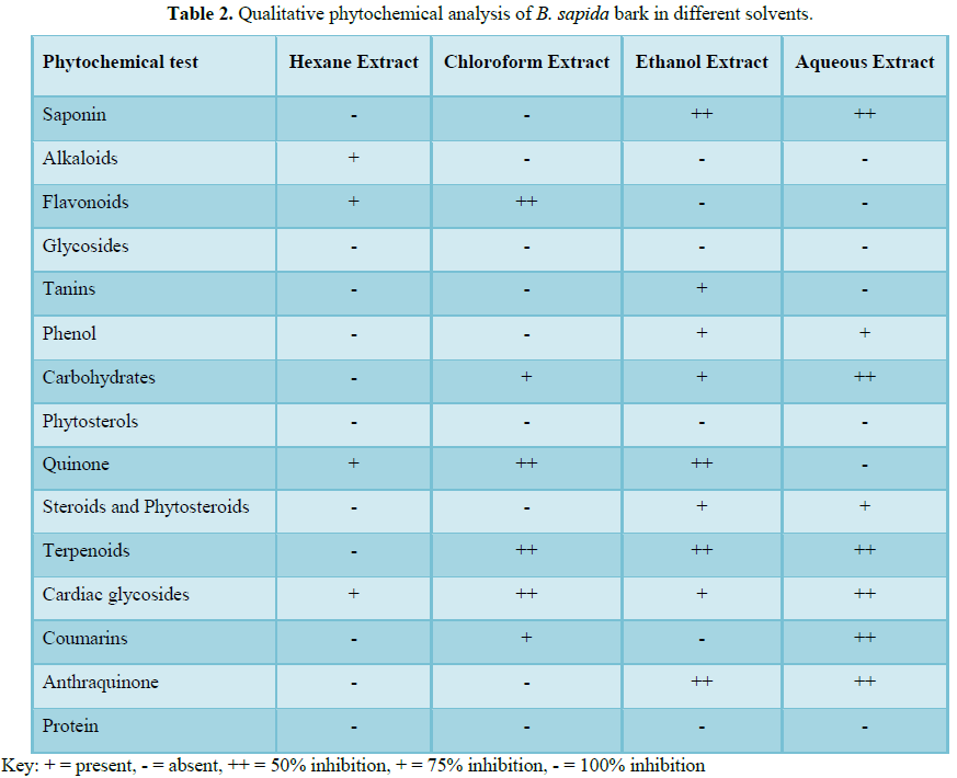
Qualitative phytochemical
analysis of B. sapida leaf and bark
in different solvents
On the dried leaf and bark samples of B. sapida collected, qualitative phytochemical screening was
carried out using ethanol, chloroform, water and N-hexane as solvents. The
following phyto-constituents such as saponin, alkaloids, flavonoids,
glycosides, tanins, phenol, carbohydrates, phytosterols, quinone, steroids,
phytosteroids, terpenoids, cardiac glycosides, coumarins, anthraquinone and
protein were extracted to varying degree.
Alkaloids saponins, tannins, phlobatannins, flavonoids, terpenes,
cardiac glycosides and combined anthraquinones were detected in the leaf of B. sapida, however, only saponins,
flavonoids, combined anthraquinones and cardiac glycosides were detected in the
stem bark (Tables 1 and 2).
Table 3 shows the percentage yield of extract in
each solvent used to ferment the extract. It was observed that leaf of the
plant had the highest yield of 5.2% using hexane for extraction and the lowest
yield of 0.4% using chloroform for extraction while the highest yield of 0.7%
and lowest yield of 0.3% was obtained for the plant bark using Hexane and
chloroform respectively for extraction.
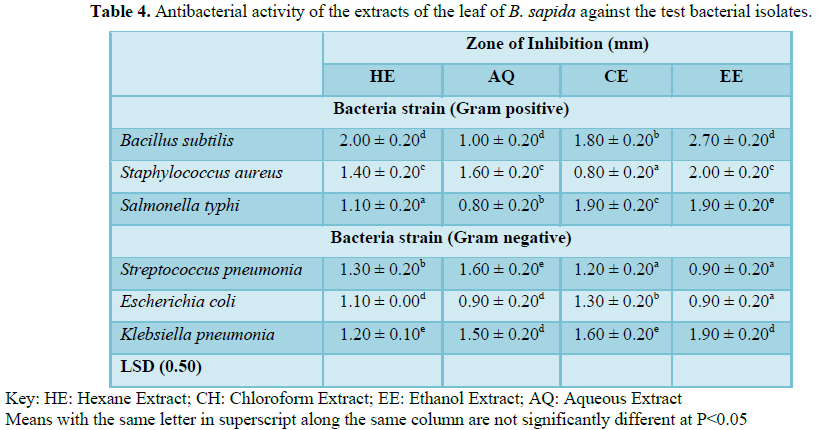
Antibacterial activity of the
extracts of stem bark of B. sapida
against the test bacterial isolates
The antibacterial activities of the stem bark extract of B. sapida and streptomycin are presented
the Table 5. The stem bark extract
inhibited the growth of B. subtilis
at concentrations 60 mg/ml and higher whereas it inhibited the growth of S. aureus only at 100 and 200 mg/ml. No
inhibition was observed for E. coli
and S. dysenteriae at the various
test concentrations.
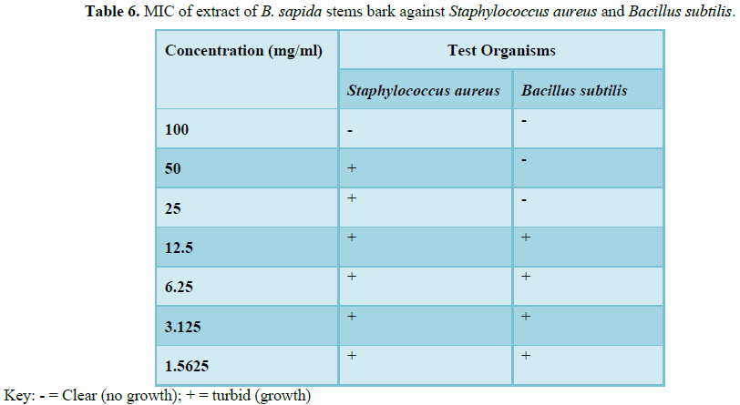
The Minimum Inhibitory
Concentration (MIC) of the stem barks extract of B. sapida for susceptible bacterial species. The MIC values
obtained were 12.5 mg/ml and 100 mg/ml for B.
subtilis and S. aureus,
respectively (Table 6).
DISCUSSION
The antimicrobial activities of B.
sapida against selected microorganisms were investigated in this study.
Form the results obtained from the qualitative phytochemical screening of B. sapida using ethanol, chloroform,
water and N-hexane as solvents for extraction, result showed that
phytochemicals such as alkaloids saponins, tannins, phlobatannins, flavonoids,
terpenes, cardiac glycosides and combined anthraquinones were detected in the
leaf of B. sapida, however, only
saponins, flavonoids, combined anthraquinones and cardiac glycosides were
detected in the stem bark. The antimicrobial activities reported in this
research could be attributed to the presence of these phytochemical
constituents of the plant. It is interesting to note that fewer phytochemical
constituents were detected in the stem bark extract than the leaf extract, yet
it performed better than the leaf extract. The stem bark of the plant demonstrating
high inhibitory activity is in agreement with the report of Sukumar et al. [13]
that the activity of phytochemical compounds on target species varies with
respect to plant parts from which they are extracted. The stem bark extracts
demonstrating higher inhibitory activities than the leaf extract could be as a
result of the phytoconstituents in the leaf were present in trace amount or
could be different in type compared to those detected in the stem bark. For
instance, saponins often occur as complex mixtures and according to the
structure of the aglycone or sapogenin, two kinds of saponins are recognized,
the steroidal and the pentacyclic type [9]. Also, there are various classes of
alkaloids, but basic nitrogen is the unifying factor, each phytoconstituent is
a group of compounds, each compound differing in structure and chemical
properties [9,14].
The results of antimicrobial sensitivity test revealed that there was
no activity against the tested fungal species at all concentration of the
extracts used. This indicates that the fungal species used in this study
demonstrated physiological resistance to the leaf and stem bark extracts of B. sapida. The absence of antifungal
activity using the extracts agrees with the report of Duraipandiyan et al.
[15], who documented that ethanolic leaf extract of B. sapida had no antifungal activity against C. albicans. However, this finding disagrees with the finding of
Nascimento et al. [2] who reported the susceptibility of C. albicans to extracts from basil, clove, guava, jambolan, lemon
balm, pomegranate, rosemary and thyme. This difference could be as a result of
the variation in the phytochemical constituents of the different plants used in
the various study.
From the results obtained from this study, it was observed that the
ethanolic leaf and stem bark extracts of B.
sapida demonstrated inhibitory activity against Gram positive bacteria (S. aureus and B. subtilis). Farjana et al. [16] also reported that ethanol
extracts of guava leaf showed antibacterial activity against S. aureus and Staphylococcus epidermidis in their study. However, the Gram
negative bacteria (E. coli and S. dysenteriae) were resistant to the
two extracts. In agreement to this finding is the report of Nascimento et al.
[2] who also reported that E. coli is
resistant to all the extracts gotten from plants used in their study. Also,
previous researches have reported E. coli
to have multi-resistance against drugs. Resistance of Gram negative bacteria to
these plant extracts could be as a result of the possession of sophisticated
cell wall by the bacteria which does not allow permeation of external agents
[17].
The plant extracts, though active to some extent against the Gram
positive bacterial species used in the study but was not as active when compare
with the effect of streptomycin, the standard drug used. Streptomycin is a
broad spectrum antibiotic, which is active against both Gram positive and Gram
negative bacteria. The lower inhibitory activity of the extracts when compared
with the standard drug may be attributed to the fact that the extracts used
were in their crude form. Also, the active phytochemical constituents of the
plant extracts acting against the bacteria could be present in trace amount
while the active constituents of streptomycin could be present in very high
amount. It is anticipated that better results could be obtained with purified
fractions of the extracts.
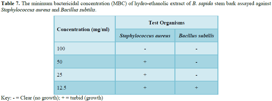
Statistical analysis revealed significant difference
(P<0.05) between the potency of leaf and stem bark extracts of B. sapida against bacteria with the stem
bark extract being more potent. Only the minimum inhibitory concentration (MIC)
of the stem bark was determined, because of aforementioned results earlier
obtained. In coincidence with the negative effect of the extract on Gram
negative bacteria. Sharma et al. [18] reported that the antidiarrheal activity
of aqueous and ethanolic stem bark extracts of B. sapida was as a result of the ability of the extracts to inhibit
intestinal motility and enter pooling effect. This implies that the usefulness
of the leaf and stem bark extracts of B.
sapida in the treatment of dysentery in folklore is not due to its
antibacterial effect on toxin producing bacteria that are associated with
diarrhea, such as E. coli and S. dysenteriae (Table 7).
CONCLUSION
This research has revealed that Blighia
sapida extracts showed antimicrobial activity against the tested
microorganisms at varying levels. Both plant parts could be the best form of
treatment to reduce the prevalence of infections caused by microorganisms. In
addition, more research can be carried out on this plant to know the most
active constituents of the plant responsible for antimicrobial activity; these
active constituents can be isolated to develop new drugs which can be used for
treatment of infections caused by microbes. Therefore being able to identify
plants and their active constituents that are potent against microorganism will
be a break through to solving the problem of emergence of resistance by
microorganisms to antibiotics as new drugs could be formulated.
CONFLICT OF INTEREST
We declare that we have no conflict of Interest.
1. Cohen
ML (1992) Epidemiology of drug resistance: implications for a
post-antimicrobial era. Science 257: 1050-1055.
2. Nascimento
GGF, Locatelli J, Freitas PC, Silva GL (2000) Antibacterial activity of plant
extracts and phytochemicals on antibiotic-resistant bacteria. Braz J Microbiol
31: 247-256.
3. Farjana
A, Zerin N, Kabir MS (2014) Antimicrobial activity of medicinal plant leaf
extracts against pathogenic bacteria. Asian Pac J Trop Dis 4: S920-S923.
4. Shihabudeen
MH, Priscilla DH, Thirumurugan K (2010) Antimicrobial activity and
phytochemical analysis of selected Indian folk medicinal plants. Int J Pharma
Sci Res 1: 430-434.
5. Sarmiento
WC, Maramba CC, Gonzales MLM (2011) An in vitro study on the antibacterial effect
of neem (Azadirachta indica) leaf
extract on methicillin-sensitive and methicillin-resistant Staphylococcus aureus. PIDSP J 12: 40-45.
6. Mukhtar
H, Ahmed N (2000) Tea polyphenols: Prevention of cancer and optimizing health.
Am J Clin Nutr 71: 1698-1702.
7. Richard
FT, Joshua AT, Phillips AJ (2013) Effect of aqueous extract of leaf and bark of
guava (Psidium guajava) on fungi Microsporum gypseum and Trichophyton mentagrophytes and bacteria
Staphylococcus aureus sand Staphylococcus epidermidis. Adv Med Plant
Res 1: 45-48.
8. Okogun
JI (1996) Medicinal plant research in Nigeria. Chem Nigerian Med Plants 10:
31-45.
9. Evans
WC (2000) Trease and Evans Pharmacognosy (15th Edn). W.B. Saunders
Company Ltd., pp: 135-150.
10. Sofowora
A (2001) Medicinal plants and traditional medicine in Africa. J Phytochem 34:
223-230.
11. Cheesebrough
M (2000) District laboratory practice in tropical African countries. Cambridge
University Press, London.
12. Akinyemi
KO, Oladapo O, Okwara CE, Ibe CC, Fasure AK (2005) Screening of crude extracts
of some medicinal plants used in South-West Nigerian Unorthodox medicine for
anti-methicilin resistant Staphylococcus
aureus. BMC Complement Altern Med 5: 6.
13. Sukumar
K, Perich MJ, Boobar LR (1991) Botanical derivatives in mosquito control: A
review. J Am Mosq Control Assoc 7: 210-237.
14. Sofowora
A (1993) Medicinal plants and traditional medicine in Africa. Spectrum Books
Ltd., Ibadan, Nigeria, pp: 191-289.
15. Duraipandiyan
V, Ayyaner M, Ignacimuthus S (2006) Antimicrobial activity of some
ethnomedicinal plants used by Paliyar tribe from Tamil Nadu, India. BMC
Complement Altern Med 6: 35.
16. Farjana
A, Zerin N, Kabi S (2014) Antimicrobial activity of medicinal plant leaf
extracts against pathogenic bacteria. Asian Pac J Trop Dis 4: 920-923.
17. Brown
WL (1975) Contributions toward a reclassification of the formicidae, V.
ponerinae, tribes Platythyreini, Cerepacchyini, Cylindromyrmecini,
Acanthostichini and Aenictogitini. Cornell University 5: 1-116.
18. Sharma
DK, Gupta VK, Kumar S, Joshi V, Mandal RS, et al. (2015) Evaluation of
antidiarrheal activity of ethanolic extract of Holarrhena antidysenterica seeds in rat. Vet World 8: 1392-1395.
-
 Table 1
 -
Table 2
-
Table 3
-
Table 4
-
Table 5
-
Table 6
-
Table 7


QUICK LINKS
- SUBMIT MANUSCRIPT
- RECOMMEND THE JOURNAL
-
SUBSCRIBE FOR ALERTS
RELATED JOURNALS
- Journal of Agriculture and Forest Meteorology Research (ISSN:2642-0449)
- Proteomics and Bioinformatics (ISSN:2641-7561)
- Journal of Microbiology and Microbial Infections (ISSN: 2689-7660)
- Journal of Veterinary and Marine Sciences (ISSN: 2689-7830)
- Journal of Astronomy and Space Research
- Food and Nutrition-Current Research (ISSN:2638-1095)
- Journal of Biochemistry and Molecular Medicine (ISSN:2641-6948)


