913
Views & Citations10
Likes & Shares
WARBURGS
HYPOTHESIS
Warburgs [1-3] findings on the origin
of cancer cells was based on the impairment in the respiratory metabolism of
normal cells. According to his hypothesis normal cells when deprived off thirty
three percent of their oxygen requirement will turn into cancer cells by
switching over to anaerobic glycolysis and the production of lactic acid. The
production of lactic acid is a specific diagnostic feature of all cancer cells
of any type of cancer and also the cells of all cancer types [1-3]. In fact
visualized that the metabolic switch in respiratory cancer cell’s aerobic
metabolism from the oxidative phase to anaerobic phase may be the causative
factor for carcinogenesis.
CANCER
ETIOLOGY
However research investigations beyond
1953, consequent to the revelation of DNA molecules being the crucial regions
for the transformation of normal cells into cancer cells, proved beyond doubt
that tumorigenesis or carcinogenesis is possible only due to various mutagenic
factors. Oncological studies have revealed that multifarious chemical compounds
in the environment, back ground radiations of the environment or the radio
nuclides, microbial infections and food contaminants can effect/bring crucial
mutations in such critical genes as proto-oncogenes and tumor suppressor genes
as well as single nucleotide polymorphic changes in the down line genes of the
core critical genes. The cumulative effects of such mutations are only
attributed as the cause for cancer cells formation [2].
CANCER
METABOLISM
The very long latent period that the
cancer disease takes for its manifestation (15-20 years), stand as evidence to
document the above accumulation of changes or mutations in the base composition
of DNA or genes. The fermentation process of energy production in cancer or the
Warburg effect may represent one of the after effects of transformation in
their metabolism and may not be considered as the etiologic reason for origin
of cancer cells or the transformation of normal cells into cancer cells. In
this context, it is of interest to note that muscle cells of all higher
organisms suffer a small bout of anoxic condition at times of hyper activity
and create an oxygen debt by the production of the fatigue poison namely the
lactic acid. However these muscle cells reinstate their normal condition as and
when the O2 supply is restored. It is also a functional adaptation
of all cancer cells to thrive with a minimal energy production through Warburg
effect. Recent studies have revealed that cancer cells maintain a micro
environmental niche wherein several pro-inflammatory cytokines/proteins, growth
factors synthesized from within as well as obtained from without enable their
survival and aberrant growth through proliferation.
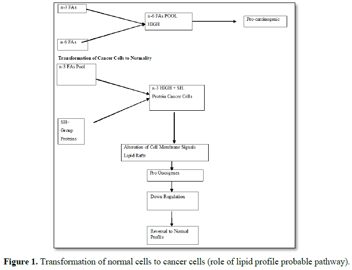
In this context, researchers optimism
that absence of sulfhydryl groups and a fatty acid partner produce a low oxygen
environment and encourage cancer cells to proliferate may be indicating the
metabolic profile of cancer cells. His view gives a cue that all normal cells
undergo some changes at the plasma membrane level and thus transform into a
malignant form where in the membrane compounds play a crucial metabolic role as
well as an immunological unresponsive potential to the host immune
surveillance.
FATTY
ACIDS DILEMMA
In our school of oncological research and thought,
it has been construed that different essential fatty acids play a role either
in enhancing or suppressing cancer as visualized previously. For instance the
synthesis of prostaglandins of D, E and F series/through arachidonic acid
induced enzyme pathway and eicosanoids and the tilt of balance between n-3 and
n-6 PUFAs towards more n-6 categories may be attributed to increased cancer risks.
However n-3 PUFA excerts anticancer effects contrary to n-6 fatty acids. In
this line, oleic acid kills breast cancer cells [8]
It has also been reported that olive oil represses
the gene viz., HER2/neu/erb-B2 involved in the development of breast cancer. By
cutting the HER2/neu by 46%, it significantly down regulates its expression.
Our personal observation on the fatty acid com position of breast cancer tissue
also revealed higher percentage of n-6 linoleic acid (24.534%) as compared to
the linolenicn-3 (0.1566%) acid [4,9]. Similar to linoleic (n-6) palmitic acid
(n-6) boosts the cancer cells survival. Our personal observation revealed
higher percentage of palmitic (n-6) similar to linoleic (n-6) It has also been
revealed that palmitic acid silences the fatty acid synthetase-Fas genes in
cancer cells and induce BRCA I expression.
Next in the line of cancer promotors is the
myristic acid. All these findings correlate that structural composition of
normal cells and the membrane components in regard to PUFA are altered to
promote carcinogenesis supporting the concept of Warburg mentioned per-se.
Recently researchers have revealed that n-3 PUFA alters cancer cell membrane’s
signaling assemblies or the lipid rafts and it represents a crucial event in down
regulating to pro-carcinogenic cell signaling and to revoke the
immunocompetence. In cancer cells several signal transducing pathway are
operated by the gain of gene signatures and the hormone responsive cancer cells
operate these pathways and well survive through their hormone specific
receptors. Thus targeting cancer cell membranes and their lipid profile with
more n-3 PUFAS seems to be a novel therapeutic approach towards cancer cure.
Such novel approach viz., membrane lipid therapy and/or Lipidomic therapy was
propounded very recently by several researchers [6,7]. These investigators have
focused such membrane based anticancer lipidomic therapy. In making this
realization pharmacopeia/pharmacognancy should involve in drug designs by
adding the anti-carcinogenic n-3 essential Fatty acids sand deleting the
innocuous n-6 FAs, Moreover the target focus towards cancer cells by such lipid
modulation, with receptor orientation is all the more beneficial and
efficacious.
n-6
VERSUS n-3 FATTY ACIDS
Considering the fact that more n-6 Fatty acids as
compared to n-3 Fatty acids and the tilt of balance towards n-6 may be
attributed to breast cancer induction, the ratio of two n-6 Fatty acids
palmitic and linoleic combined percentage with one n-3linolenicrevealed ratio
of 1:50, respectively in the present study. Though another n-3 oleic acid was
in high proportion its ratio with n-6 palmitic and linoleic was 1:1.5,
respectively.
To confirm whether this higher percentage n-6
linoleic-palmitic as compared to linolenic is specific to breast cancer alone
or is a generalized and common factor to other types of cancers, the percentage
difference of them in the colorectal and uterine- cervical cancerous tissues
were observed [10,11]. In the uterine cervical tissues the palmitic and
linoleic percentage was found to be 8.36 and 60.80, respectively. The linolenic
was found in the range 0.29 to 0.55%. In the same tissue the methylated
palmitate showed a range of 4.2 to 14.0%. While the methyl linoleate showed a
range of 1.19 to 1.93% and the cholesterol showed a range between 47.5% to 85%.
The linolenic was negligibly in low percentage of 0.29. In our study on free
fatty acid analysis of three different cancerous tissues (breast,
gastro-intestine, uterine-cervical) the short chain fatty acid namely the
butyric acid which is the end product of microbial digestion of carbohydrates
is conspicuously absent. Considering the multifarious functions of butyric acid
viz., induction of apoptosis; cell cycle arrest; inhibition of histone deacetylases;
regulation of aromatase promoters; alter the mitochondrial membrane potential,
enhances apoptosis in cancers by a Ca++ dependent
mitochondrial-intrinsic pathway, promotes FAS mediated cancer cell apoptosis;
activates caspace-8, the conspicuous absence of butyric acid alongside n-6
fatty acids increase may be taken as evidence that these changes in Fatty acids
profile are strong prognostic indicators for cancer development.
LONG
CHAIN FATTY ACIDS AND CANCER
In recent years malignancy of cancer has been
correlated to fatty acid metabolism. Since all cancers and especially the
breast, uterus, ovary, cervical and all sub site cancers of gastro intestinal
tract express hormone receptors viz., estrogen, progesterone, etc., it is
postulated that fat content of these tissues particularly cholesterol may act
as a precursor for the synthesis of above hormones.
CHOLESTEROL
AND CANCER
In our study on uterine-cervical cancer tissue,
the content of cholesterol was found predominantly in higher percentage. Harn
et al. [12] have revealed that endometrial hyperplasia and endometrial in situ
carcinoma have been ascribed to the cholesterol derived hormone estrogen.
Recently investigator at Simon Frasher University have revealed that
cholesterol binding puts the brakes on oxosterol related proteins ability to
couple phosphatidyl inositol 4-phosphate and accelerates cell growth crazy.
These cholesterol may function as an enhancer of cellular metaplasia.
OLEIC
ACID - THERAPEUTIC SIGNIFICANCE
Oleic acid is a fatty acid that occurs naturally
in various animal and vegetable fats and oils. Chemically it is CH3(CH2)7CH=CH(CH2)7COOH.
In the present study the significance of a markedly high percentage of oleic
acid could not be explained, in view of the report that it kills cancer cells
[8]. However the above result is of significance and therapeutic value in view
of another observation that oleic acid boosts the effectiveness of the
anti-cancer drug herceptin and helped to prolong the lives of many cancer
patients.
THERAPEUTIC
SIGNIFICANCE AND SELECTION
In the present study the fatty acid composition of
normal breast tissue could not be reported due to difficulty in procuring the
control samples from female subjects. However the present results are compared,
taking in to account the reports of previous investigations. The serious draw
back in such comparison may not also be unexpected. Inside the breast tissues
of normal subjects the level of fatty acids belonging to both n-3 and n-6
categories could be in a state of flux, depending upon the various
physiological and gynecological conditions of the female subjects. Holmes [13]
in their paper revealed that there was no association of breast cancer
incidence in both the pre-menopausal and post-menopausal groups (cohort study)
with intake of animal fat, vegetables fat, polyunsaturated fat, saturated fat
or cholesterol. In their paper they have reported that certain fatty acids like
(n6-linoleic, Palmitic or PUFA) have modulated mammary tumors growth and
metastasis in animals, while omega-3 from marine origin is endowed with an
inhibitory effect. These authors also cited that human ecological studies
supported the above observation [14-18]. However in an established and
differentiated cancer tissue of breast the above flux may not be expected but
only the tilt of balance between n-3 and n-6 FAs towards n-6 FAs, as the latter
has been obviously attributed to promote the carcinogenesis. Hence what is
presented in our present observation and the uniform pattern of the EFAs, (i)
higher percentage of linoleic acid and palmitic acid; (ii) the complete absence
of butyric acid, the short chain fatty acid; (iii) Higher percentage of
cholesterol and iv. the negligible percentage (<1%) of linolenic acid in the
different cancer tissues (stomach, colon, rectum, uterine-cervix, mammary,
breast) may be considered as important bio markers to decide upon the mode of
treatment procedures and also the phytonutritional requirements as adjuvants to
prolong the survival period as well as to build up immunity which has been
deprived by the cancer growth and proliferation.
1.
Warburg O (1928) The chemical constitution of respiration
ferment. Science 68: 437-443.
2.
Warburg O (1956) On respiratory impairment in cancer
cells. Science 124: 269-270.
3.
Warburg O (1956) On the origin of cancer cells. Science
123: 309-314.
4.
Ramalingam K (2015) Gene signatures and gene function
profiles in cancer manifestation - A review. Int J Recent Sci Res 6: 3254-3258.
5.
Pattersew CHH (2012) The effect of oxega-3
polyunsaturated fatty acids on human cancer cells. Ph.D. Thesis, Norwegian
university of science and technology. Faculty of Medicine Trondheim. Norway.
6.
Escriba PV, Busquets X, Inokuchi J, Balogh G, Torok Z, et
al. (2015) Membrane lipid therapy: modulation of the cell membrane composition
and structure as a molecular base for drug discovery and new disease treatment.
Prog Lipid Res 59: 38-53.
7.
Zabla S, Hagen TLM (2017) Cell membrane modulation as
adjuvant in cancer therapy. Cancer Treat Rev 52: 48-57.
8.
Carillo CM, Caviadel M, Alonso-Torre SR (2012) Anti-tumor
effect of oleic acid: mechanisms of action: A review. Nutr Hosp 27: 1860-1865.
9.
Kanakavalli S (2011) Studies on the epidemiology,
histopathology and genomic details of breast cancer in South Indian women with
supplementary cell line observations and mathematical modeling.
10.
Rajeswari T (2011) Studies on epidemiology,
histopathology and genetic details of uterine cervical cancer in South Indian
women with supplementary cell line observations and statistical modeling.
University of Madras. Ph. D. Thesis.
11.
Inda Alias Muthu Meena P (2013) Studies on epidemiology,
histopathology and genetic details of gastrointestinal cancer in Tamil Nadu
with supplementary cell line observations and statistical modeling: Human
disease related immune biotechnological approaches. Mother Teresa Women’s
University. Ph. D. Thesis.
12.
Harn N, Allan C, Oliver C, Middaugh CR (2007) Highly
concentrated monoclonal antibody solutions. Direct analysis of physical
structure and thermal stability. J Pharm Sci 96: 532-546.
13.
Holmes D (2012) Association of dietary intake of fat and
fatty acids with risk of breast cancer. JAMA 281: 1-10.
14.
Hanahan D, weinberg RA (2011) Hallmarks of cancer. The
Next Generation Cell 144: 646-674.
15.
Hurseiw JS (2013) Cell membrane fatty acids and health. J
Pharm Sci 5: 38-46.
16.
Pivnyuk MVF, Chekhu (2013) Alternation in lipid
composition of plasma membrane of sensitive and resistant Guerin carcinoma
cells due to action of free liposomal form of cisplatin L.A. Exp Oncol 35:
192-197.
17.
Ramalingam K (2016) Cytomorphology of gastrointestinal
cancers of South Indian cohort - Case studies report. Acta Biomedica Scientia
3: 102-108.
18.
Veronique C, Marie C, Karen (2006) Acetyle co A
carboxylase is essential to breast cancer survival cancer Pres 66: 5287-5294.
-
 Table 1

QUICK LINKS
- SUBMIT MANUSCRIPT
- RECOMMEND THE JOURNAL
-
SUBSCRIBE FOR ALERTS
RELATED JOURNALS
- International Journal of Radiography Imaging & Radiation Therapy (ISSN:2642-0392)
- Journal of Psychiatry and Psychology Research (ISSN:2640-6136)
- Journal of Ageing and Restorative Medicine (ISSN:2637-7403)
- Journal of Rheumatology Research (ISSN:2641-6999)
- Journal of Carcinogenesis and Mutagenesis Research (ISSN: 2643-0541)
- International Journal of Diabetes (ISSN: 2644-3031)
- Journal of Allergy Research (ISSN:2642-326X)


