1139
Views & Citations139
Likes & Shares
External dental
fistula often presents with facial sinus, and patients visit dermatology
clinics. Usually, diagnosis is not
difficult by orthopantomography. We present a case of external dental fistula
which was not detected by orthopantomography, but examination by cone-beam
computed tomography (CT) revealed a small sequestrum.
CASE REPORT
A 62-year-old woman was referred
to our hospital, complaining of a nodular lesion on her right cheek, with 2
months’ duration. Previously, she
visited otolaryngology department, and needle biopsy showed no malignancy. Administration of antibiotics resulted in no
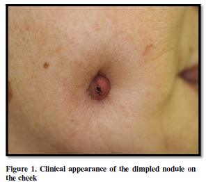
effects. On the initial visit to our
department, a physical examination showed a reddish dimpled nodule located on
the right cheek (Figure 1). Clinical diagnosis was external dental fistula,
however, examination by pantomography did not detect any abnormalities. Histological examination showed non-specific
granulation with dense infiltration of inflammatory cells composed of
lymphocytes, neutrophils, histiocytes and plasma cells in the whole dermis (Figure
2). Tissue cultures for bacteria, mycobacterium tuberculosis and
non-tuberculous mycobacterium were all sterile. Also, polymerase chain reaction
(RCR) analysis for mycobacterium tuberculosis and non-tuberculous mycobacterium
were negative. Laboratory examination
showed no abnormalities including liver and kidney function, and tuberculin
test revealed negative reaction.
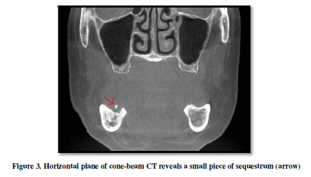
Cone-beam computed tomography revealed the presence of a small
sequestrum (Figure 3). Surgical
treatment with sequestrectomy resulted in improvement with scar within 3 months.
DISCUSSION
REFERENCES
1. Wilson SW, Ward DJ, Burns A (2001) Dental infections masquerading as
skin lesions. Br J Plast Surg 54: 358-360. PMID: 11355994
2. Patel S, Kanagasingam S, Mannocci F (2010) Cone beam computed
tomography (CBCT) in endodontics. Dent Update 37: 373-379. PMID: 20929151

QUICK LINKS
- SUBMIT MANUSCRIPT
- RECOMMEND THE JOURNAL
-
SUBSCRIBE FOR ALERTS
RELATED JOURNALS
- International Journal of Surgery and Invasive Procedures (ISSN:2640-0820)
- Journal of Clinical Trials and Research (ISSN:2637-7373)
- Oncology Clinics and Research (ISSN: 2643-055X)
- Ophthalmology Clinics and Research (ISSN:2638-115X)
- Journal of Renal Transplantation Science (ISSN:2640-0847)
- Journal of Alcoholism Clinical Research
- Journal of Forensic Research and Criminal Investigation (ISSN: 2640-0846)


